| The transverse process of the atlas is about 1 cm. below and in front of the apex of the mastoid process. The transverse process of the sixth cervical vertebra is opposite the cricoid cartilage; below it is the transverse process of the seventh and occasionally a cervical rib. | 2 |
 |
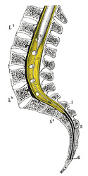
FIG. 1213– Sagittal section of vertebral canal to show the lower end of the medulla spinalis and the flum terminale. (Testut.) Li, Lv. First and fifth lumbar vertebra. S\??\ Second sacral vertebra. 1. Dura mater. 2. Lower part of subarachnoid cavity. 3. Lower extremity of medulla spinalis. 4. Filum terminale internum, and 5. Filum terminale externum. 6. Attachment of filum terminale to first segment of cooccyx. (See enlarged image) |
| |
 |
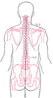
FIG. 1214– Scheme showing the relations of the regions of attachment of the spinal nerves to the vertebral spinous processes. (After Reid.) (See enlarged image) |
| |
| |
| Medulla Spinalis.—The position of the lower end of the medulla spinalis varies slightly with the movements of the vertebral column, but, in the adult, in the upright posture it is usually at the level of the spinous process of the second lumbar vertebra (Fig. 1212); at birth it lies at the level of the fourth lumbar. | 3 |
| The subdural and subarachnoid cavities end below opposite the spinous process of the third sacral vertebra (Fig. 1213). | 4 |
| |
| Spinal Nerves (Fig. 1214).—The table on page 1305, after Macalister, shows the relations which the places of attachment of the nerves to the medulla spinalis present to the bodies and spinous processes of the vertebræ. | 5 |


