(Articulatio Genu)
The knee-joint was formerly described as a ginglymus or hinge-joint, but is really of a much more complicated character. It must be regarded as consisting of three articulations in one: two condyloid joints, one between each condyle of the femur and the corresponding meniscus and condyle of the tibia; and a third between the patella and the femur, partly arthrodial, but not completely so, since the articular surfaces are not mutually adapted to each other, so that the movement is not a simple gliding one. This view of the construction of the knee-joint receives confirmation from the study of the articulation in some of the lower mammals, where, corresponding to these three subdivisions, three synovial cavities are sometimes found, either entirely distinct or only connected together by small communications. This view is further rendered probable by the existence in the middle of the joint of the two cruciate ligaments, which must be regarded as the collateral ligaments of the medial and lateral joints. The existence of the patellar fold of synovial membrane would further indicate a tendency to separation of the synovial cavity into two minor sacs, one corresponding to the lateral and the other to the medial joint. | 1 |
| The bones are connected together by the following ligaments: | 2 |
| The Articular Capsule. |
|
The Anterior Cruciate. |
| The Ligamentum Patellæ. |
|
The Posterior Cruciate. |
| The Oblique Popliteal. |
|
The Medial and Lateral Menisci. |
| The Tibial Collateral. |
|
The Transverse. |
| The Fibular Collateral. |
|
The Coronary. |
|
| |
| The Articular Capsule (capsula articularis; capsular ligament) (Fig. 345).—The articular capsule consists of a thin, but strong, fibrous membrane which is strengthened in almost its entire extent by bands inseparably connected with it. Above and in front, beneath the tendon of the Quadriceps femoris, it is represented only by the synovial membrane. Its chief strengthening bands are derived from the fascia lata and from the tendons surrounding the joint. In front, expansions from the Vasti and from the fascia lata and its iliotibial band fill in the intervals between the anterior and collateral ligaments, constituting the medial and lateral patellar retinacula. Behind the capsule consists of vertical fibers which arise from the condyles and from the sides of the intercondyloid fossa of the femur; the posterior part of the capsule is therefore situated on the sides of and in front of the cruciate ligaments, which are thus excluded from the joint cavity. Behind the cruciate ligaments is the oblique popliteal ligament which is augmented by fibers derived from the tendon of the Semimembranosus. Laterally, a prolongation from the iliotibial band fills in the interval between the oblique popliteal and the fibular collateral ligaments, and partly covers the latter. Medially, expansions from the Sartorius and Semimembranosus pass upward to the tibial collateral ligament and strengthen the capsule. | 3 |
| |
| The Ligamentum Patellæ (anterior ligament) (Fig. 345).—The ligamentum patellæ is the central portion of the common tendon of the Quadriceps femoris, which is continued from the patella to the tuberosity of the tibia. It is a strong, flat, ligamentous band, about 8 cm. in length, attached, above, to the apex and adjoining margins of the patella and the rough depression on its posterior surface; below, to the tuberosity of the tibia; its superficial fibers are continuous over the front of the patella with those of the tendon of the Quadriceps femoris. The medial and lateral portions of the tendon of the Quadriceps pass down on either side of the patella, to be inserted into the upper extremity of the tibia on either side of the tuberosity; these portions merge into the capsule, as stated above, forming the medial and lateral patellar retinacula. The posterior surface of the ligamentum patellæ is separated from the synovial membrane of the joint by a large infrapatellar pad of fat, and from the tibia by a bursa. | 4 |
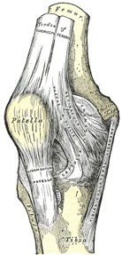 |
FIG. 345– Right knee-joint. Anterior view. (See enlarged image) |
| |
| |
| The Oblique Popliteal Ligament (ligamentum popliteum obliquum; posterior ligament) (Fig. 346).—This ligament is a broad, flat, fibrous band, formed of fasciculi separated from one another by apertures for the passage of vessels and nerves. It is attached above to the upper margin of the intercondyloid fossa and posterior surface of the femur close to the articular margins of the condyles, and below to the posterior margin of the head of the tibia. Superficial to the main part of the ligament is a strong fasciculus, derived from the tendon of the Semimembranosus and passing from the back part of the medial condyle of the tibia obliquely upward and lateralward to the back part of the lateral condyle of the femur. The oblique popliteal ligament forms part of the floor of the popliteal fossa, and the popliteal artery rests upon it. | 5 |
| |
| The Tibial Collateral Ligament (ligamentum collaterale tibiale; internal lateral ligament) (Fig. 345).—The tibial collateral is a broad, flat, membranous band, situated nearer to the back than to the front of the joint. It is attached, above, to the medial condyle of the femur immediately below the adductor tubercle; below, to the medial condyle and medial surface of the body of the tibia. The fibers of the posterior part of the ligament are short and incline backward as they descend; they are inserted into the tibia above the groove for the Semimembranosus. The anterior part of the ligament is a flattened band, about 10 cm. long, which inclines forward as it descends. It is inserted into the medial surface of the body of the tibia about 2.5 cm. below the level of the condyle. It is crossed, at its lower part, by the tendons of the Sartorius, Gracilis, and Semitendinosus, a bursa being interposed. Its deep surface covers the inferior medial genicular vessels and nerve and the anterior portion of the tendon of the Semimembranosus, with which it is connected by a few fibers; it is intimately adherent to the medial meniscus. | 6 |
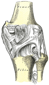 |
FIG. 346– Right knee-joint. Posterior view. (See enlarged image) |
| |
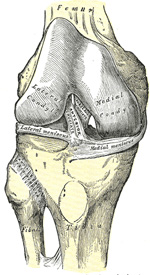 |
FIG. 347– Right knee-joint, from the front, showing interior ligaments. (See enlarged image) |
| |
| |
| The Fibular Collateral Ligament (ligamentum collaterale fibulare; external lateral or long external lateral ligament) (Fig. 348).—The fibular collateral is a strong, rounded, fibrous cord, attached, above, to the back part of the lateral condyle of the femur, immediately above the groove for the tendon of the Popliteus; below, to the lateral side of the head of the fibula, in front of the styloid process. The greater part of its lateral surface is covered by the tendon of the Biceps femoris; the tendon, however, divides at its insertion into two parts, which are separated by the ligament. Deep to the ligament are the tendon of the Popliteus, and the inferior lateral genicular vessels and nerve. The ligament has no attachment to the lateral meniscus. | 7 |
| An inconstant bundle of fibers, the short fibular collateral ligament, is placed behind and parallel with the preceding, attached, above, to the lower and back part of the lateral condyle of the femur; below, to the summit of the styloid process of the fibula. Passing deep to it are the tendon of the Popliteus, and the inferior lateral genicular vessels and nerve. | 8 |
| |
| The Cruciate Ligaments (ligamenta cruciata genu; crucial ligaments).—The cruciate ligaments are of considerable strength, situated in the middle of the joint, nearer to its posterior than to its anterior surface. They are called cruciate because they cross each other somewhat like the lines of the letter X; and have received the names anterior and posterior, from the position of their attachments to the tibia. | 9 |
| The Anterior Cruciate Ligament (ligamentum cruciatum anterius; external crucial ligament) (Fig. 347) is attached to the depression in front of the intercondyloid eminence of the tibia, being blended with the anterior extremity of the lateral meniscus; it passes upward, backward, and lateralward, and is fixed into the medial and back part of the lateral condyle of the femur. | 10 |
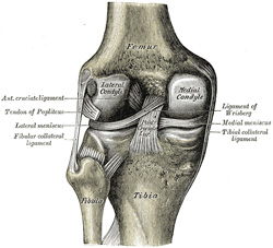 |
FIG. 348– Left knee-joint from behind, showing interior ligaments. (See enlarged image) |
| |
| The Posterior Cruciate Ligament (ligamentum cruciatum posterius; internal crucial ligament) (Fig. 348) is stronger, but shorter and less oblique in its direction, than the anterior. It is attached to the posterior intercondyloid fossa of the tibia, and to the posterior extremity of the lateral meniscus; and passes upward, forward, and medialward, to be fixed into the lateral and front part of the medial condyle of the femur. | 11 |
| |
| The Menisci (semilunar fibrocartilages) (Fig. 349).—The menisci are two crescentic lamellæ, which serve to deepen the surfaces of the head of the tibia for articulation with the condyles of the femur. The peripheral border of each meniscus is thick, convex, and attached to the inside of the capsule of the joint; the opposite border
is thin, concave, and free. The upper surfaces of the menisci are concave, and in contact with the condyles of the femur; their lower surfaces are flat, and rest upon the head of the tibia; both surfaces are smooth, and invested by synovial membrane. Each meniscus covers approximately the peripheral two-thirds of the corresponding articular surface of the tibia. | 12 |
| The medial meniscus (meniscus medialis; internal semilunar fibrocartilage) is nearly semicircular in form, a little elongated from before backward, and broader behind than in front; its anterior end, thin and pointed, is attached to the anterior intercondyloid fossa of the tibia, in front of the anterior cruciate ligament; its posterior end is fixed to the posterior intercondyloid fossa of the tibia, between the attachments of the lateral meniscus and the posterior cruciate ligament. | 13 |
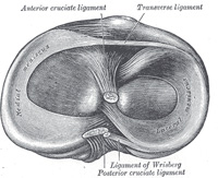 |
FIG. 349– Head of right tibia seen from above, showing menisci and attachments of ligaments. (See enlarged image) |
| |
| The lateral meniscus (meniscus lateralis; external semilunar fibrocartilage) is nearly circular and covers a larger portion of the articular surface than the medial one. It is grooved laterally for the tendon of the Popliteus, which separates it from the fibular collateral ligament. Its anterior end is attached in front of the intercondyloid eminence of the tibia, lateral to, and behind, the anterior cruciate ligament, with which it blends; the posterior end is attached behind the intercondyloid eminence of the tibia and in front of the posterior end of the medial meniscus. The anterior attachment of the lateral meniscus is twisted on itself so that its free margin looks backward and upward, its anterior end resting on a sloping shelf of bone on the front of the lateral process of the intercondyloid eminence. Close to its posterior attachment it sends off a strong fasciculus, the ligament of Wrisberg (Figs. 348, 349), which passes upward and medialward, to be inserted into the medial condyle of the femur, immediately behind the attachment of the posterior cruciate ligament. Occasionally a small fasciculus passes forward to be inserted into the lateral part of the anterior cruciate ligament. The lateral meniscus gives off from its anterior convex margin a fasciculus which forms the transverse ligament. | 14 |
| |
| The Transverse Ligament (ligamentum transversum genu).—The transverse ligament connects the anterior convex margin of the lateral meniscus to the anterior end of the medial meniscus; its thickness varies considerably in different subjects, and it is sometimes absent. | 15 |
| The coronary ligaments are merely portions of the capsule, which connect the periphery of each meniscus with the margin of the head of the tibia. | 16 |
| |
| Synovial Membrane.—The synovial membrane of the knee-joint is the largest and most extensive in the body. Commencing at the upper border of the patella, it forms a large cul-de-sac beneath the Quadriceps femoris (Figs. 350, 351) on the lower part of the front of the femur, and frequently communicates with a bursa interposed between the tendon and the front of the femur. The pouch of synovial membrane between the Quadriceps and front of the femur is supported, during the movements of the knee, by a small muscle, the Articularis genu, which is inserted into it. On either side of the patella, the synovial membrane extends beneath the aponeuroses of the Vasti, and more especially beneath that of the Vastus medialis. Below the patella it is separated from the ligamentum patellæ by a considerable quantity of fat, known as the infrapatellar pad. From the medial and lateral borders of the articular surface of the patella, reduplications of the synovial membrane project into the interior of the joint. These form two fringe-like folds termed the alar folds; below, these folds converge and are continued as a single band, the patellar fold (ligamentum mucosum), to the front of the intercondyloid fossa of the femur. On either side of the joint, the synovial membrane passes downward from the femur, lining the capsule to its point of attachment to the menisci; it may then be traced over the upper surfaces of these to their free borders, and thence along their under surfaces to the tibia (Figs. 351, 352). At the back part of the lateral meniscus it forms a cul-de-sac between the groove on its surface and the tendon of the Popliteus; it is reflected across the front of the cruciate ligaments, which are therefore situated outside the synovial cavity. | 17 |
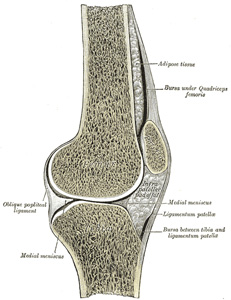 |
FIG. 350– Sagittal section of right knee-joint. (See enlarged image) |
| |
| |
| Bursæ.—The bursæ near the knee-joint are the following: In front there are four bursæ: a large one is interposed between the patella and the skin, a small one between the upper part of the tibia and the ligamentum patellæ, a third between the lower part of the tuberosity of the tibia and the skin, and a fourth between the anterior surface of the lower part of the femur and the deep surface of the Quadriceps femoris, usually communicating with the knee-joint. Laterally there are four bursæ: (1) one (which sometimes communicates with the joint) between the lateral head of the Gastrocnemius and the capsule; (2) one between the fibular collateral ligament and the tendon of the Biceps; (3) one between the fibular collateral ligament and the tendon of the Popliteus (this is sometimes only an expansion from the next bursa); (4) one between the tendon of the Popliteus and the lateral condyle of the femur, usually an extension from the synovial membrane of the joint. Medially, there are five bursæ: (1) one between the medial head of the Gastrocnemius and the capsule; this sends a prolongation between the tendon of the medial head of the Gastrocnemius and the tendon of the Semimembranosus and often communicates with the joint; (2) one superficial to the tibial collateral ligament, between it and the tendons of the Sartorius, Gracilis, and Semitendinosus; (3) one deep to the tibial collateral ligament, between it and the tendon of the Semimembranosus (this is sometimes only an expansion from the next bursa); (4) one between the tendon of the Semimembranosus and the head of the tibia; (5) occasionally there is a bursa between the tendons of the Semimembranosus and Semitendinosus. | 18 |
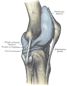 |
FIG. 351– Capsule of right knee-joint (distended). Lateral aspect. (See enlarged image) |
| |
| |
| Structures Around the Joint.—In front, and at the sides, is the Quadriceps femoris; laterally the tendons of the Biceps femoris and Popliteus and the common peroneal nerve; medially, the Sartorius, Gracilis, Semitendinosus, and Semimembranosus; behind, the popliteal vessels and the tibial nerve, Popliteus, Plantaris, and medial and lateral heads of the Gastrocnemius, some lymph glands, and fat. | 19 |
| The arteries supplying the joint are the highest genicular (anastomotica magna), a branch of the femoral, the genicular branches of the popliteal, the recurrent branches of the anterior tibial, and the descending branch from the lateral femoral circumflex of the profunda femoris. | 20 |
| The nerves are derived from the obturator, femoral, tibial, and common peroneal. | 21 |
| |
| Movements.—The movements which take place at the knee-joint are flexion and extension, and, in certain positions of the joint, internal and external rotation. The movements of flexion and extension at this joint differ from those in a typical hinge-joint, such as the elbow, in that (a) the axis around which motion takes place is not a fixed one, but shifts forward during extension and backward during flexion; (b) the commencement of flexion and the end of extension are accompanied by rotatory movements associated with the fixation of the limb in a position of great stability. The movement from full flexion to full extension may therefore be described in three phases: | 22 |
| 1. In the fully flexed condition the posterior parts of the femoral condyles rest on the corresponding portions of the meniscotibial surfaces, and in this position a slight amount of simple rolling movement is allowed. | 23 |
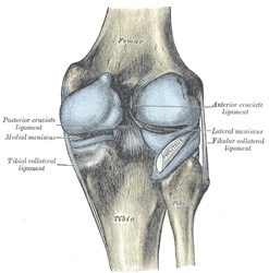 |
FIG. 352– Capsule of right knee-joint (distended). Posterior aspect. (See enlarged image) |
| |
| 2. During the passage of the limb from the flexed to the extended position a gliding movement is superposed on the rolling, so that the axis, which at the commencement is represented by a line through the inner and outer condyles of the femur, gradually shifts forward. In this part of the movement, the posterior two-thirds of the tibial articular surfaces of the two femoral condyles are involved, and as these have similar curvatures and are parallel to one another, they move forward equally. | 24 |
| 3. The lateral condyle of the femur is brought almost to rest by the tightening of the anterior cruciate ligament; it moves, however, slightly forward and medialward, pushing before it the anterior part of the lateral meniscus. The tibial surface on the medial condyle is prolonged farther forward than that on the lateral, and this prolongation is directed lateralward. When, therefore, the movement forward of the condyles is checked by the anterior cruciate ligament, continued muscular action causes the medial condyle, dragging with it the meniscus, to travel backward and medialward, thus producing an internal rotation of the thigh on the leg. When the position of full extension is reached the lateral part of the groove on the lateral condyle is pressed against the anterior part of the corresponding meniscus, while the medial part of the groove rests on the articular margin in front of the lateral process of the tibial intercondyloid eminence. Into the groove on the medial condyle is fitted the anterior part of the medial meniscus, while the anterior cruciate ligament and the articular margin in front of the medial process of the tibial intercondyloid eminence are received into the forepart of the intercondyloid fossa of the femur. This third phase by which all these parts are brought into accurate apposition is known as the “screwing home,” or locking movement of the joint. | 25 |
| The complete movement of flexion is the converse of that described above, and is therefore preceded by an external rotation of the femur which unlocks the extended joint. | 26 |
| The axes around which the movements of flexion and extension take place are not precisely at right angles to either bone; in flexion, the femur and tibia are in the same plane, but in extension the one bone forms an angle, opening lateralward with the other. | 27 |
| In addition to the rotatory movements associated with the completion of extension and the initiation of flexion, rotation inward or outward can be effected when the joint is partially flexed; these movements take place mainly between the tibia and the menisci, and are freest when the leg is bent at right angles with the thigh. | 28 |
| Movements of Patella.—The articular surface of the patella is indistinctly divided into seven facets—upper, middle, and lower horizontal pairs, and a medial perpendicular facet (Fig. 353). When the knee is forcibly flexed, the medial perpendicular facet is in contact with the semilunar surface on the lateral part of the medial condyle; this semilunar surface is a prolongation backward of the medial part of the patellar surface. As the leg is carried from the flexed to the extended position, first the highest pair, then the middle pair, and lastly the lowest pair of horizontal facets is successively brought into contact with the patellar surface of the femur. In the extended position, when the Quadriceps femoris is relaxed, the patella lies loosely on the front of the lower end of the femur. | 29 |
| During flexion, the ligamentum patellæ is put upon the stretch, and in extreme flexion the posterior cruciate ligament, the oblique popliteal, and collateral ligaments, and, to a slight extent, the anterior cruciate ligament, are relaxed. Flexion is checked during life by the contact of the leg with the thigh. When the knee-joint is fully extended the oblique popliteal and collateral ligaments, the anterior cruciate ligament, and the posterior cruciate ligament, are rendered tense; in the act of extending the knee, the ligamentum patellæ is tightened by the Quadriceps femoris, but in full extension with the heel supported it is relaxed. Rotation inward is checked by the anterior cruciate ligament; rotation outward tends to uncross and relax the cruciate ligaments, but is checked by the tibial collateral ligament. The main function of the cruciate ligament is to act as a direct bond between the tibia and femur and to prevent the former bone from being carried too far backward or forward. They also assist the collateral ligaments in resisting any bending of the joint to either side. The menisci are intended, as it seems, to adapt the surfaces of the tibia to the shape of the femoral condyles to a certain extent, so as to fill up the intervals which would otherwise be left in the varying positions of the joint, and to obviate the jars which would be so frequently transmitted up the limb in jumping or by falls on the feet; also to permit of the two varieties of motion, flexion and extension, and rotation, as explained above. The patella is a great defence to the front of the knee-joint, and distributes upon a large and tolerably even surface, during kneeling, the pressure which would otherwise fall upon the prominent ridges of the condyles; it also affords leverage to the Quadriceps femoris. | 30 |
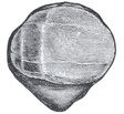 |
FIG. 353– Posterior surface of the right patella, showing diagrammatically the areas of contact with the femur in different positions of the knee. (See enlarged image) |
| |
| When standing erect in the attitude of “attention,” the weight of the body falls in front of a line carried across the centers of the knee-joints, and therefore tends to produce overextension of the articulations; this, however, is prevented by the tension of the anterior cruciate, oblique popliteal, and collateral ligaments. | 31 |
| Extension of the leg on the thigh is performed by the Quadriceps femoris; flexion by the Biceps femoris, Semitendinosus, and Semimembranosus, assisted by the Gracilis, Sartorius, Gastrocnemius, Popliteus, and Plantaris. Rotation outward is effected by the Biceps femoris, and rotation inward by the Popliteus, Semitendinosus, and, to a slight extent, the Semimembranosus, the Sartorius, and the Gracilis. The Popliteus comes into action especially at the commencement of the movement of flexion of the knee; by its contraction the leg is rotated inward, or, if the tibia be fixed, the thigh is rotated outward, and the knee-joint is unlocked. | 32 |


