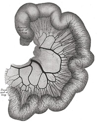
FIG. 535– Loop of small intestine showing distribution of intestinal arteries. (From a preparation by Mr. Hamilton Drummond.) The vessels were injected while the gut was in situ; the gut was then removed, and an x-ray photograph taken. (See enlarged image)
|