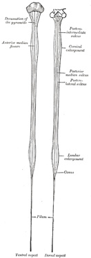
Enlargements.—The medulla spinalis is not quite cylindrical, being slightly flattened from before backward; it also
| Anatomy of the Human Body > Page 751 | CONTENTS · ILLUSTRATIONS · SUBJECT INDEX |
| PREVIOUS | NEXT | ||
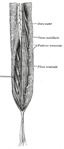
| by, and is adherent to, the dura mater; it extends downward from the apex of the tubular sheath and is attached to the back of the first segment of the coccyx. The filum terminale consists mainly of fibrous tissue, continuous above with that of the pia mater. Adhering to its outer surface, however, are a few strands of nerve fibers which probably represent rudimentary second and third coccygeal nerves; further, the central canal of the medulla spinalis extends downward into it for 5 or 6 cm. |
|
|
Enlargements.—The medulla spinalis is not quite cylindrical, being slightly flattened from before backward; it also |
| PREVIOUS | NEXT |