(Aorta Abdominalis)
The abdominal aorta (Fig. 531) begins at the aortic hiatus of the diaphragm, in front of the lower border of the body of the last thoracic vertebra, and, descending in front of the vertebral column, ends on the body of the fourth lumbar vertebra, commonly a little to the left of the middle line, 103 by dividing into the two common iliac arteries. It diminishes rapidly in size, in consequence of the many large branches which it gives off. As it lies upon the bodies of the vertebræ, the curve which it describes is convex forward, the summit of the convexity corresponding to the third lumbar vertebra. | 1 |
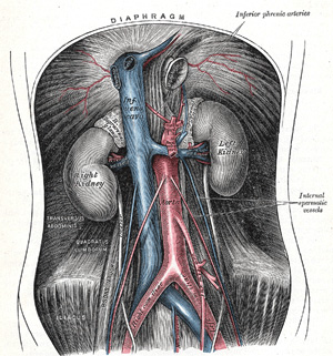 |
FIG. 531– The abdominal aorta and its branches. (See enlarged image) |
| |
| |
| Relations.—The abdominal aorta is covered, anteriorly, by the lesser omentum and stomach, behind which are the branches of the celiac artery and the celiac plexus; below these, by the lienal vein, the pancreas, the left renal vein, the inferior part of the duodenum, the mesentery, and aortic plexus. Posteriorly, it is separated from the lumbar vertebræ and intervertebral fibrocartilages by the anterior longitudinal ligament and left lumbar veins. On the right side it is in relation above with the azygos vein, cisterna chyli, thoracic duct, and the right crus of the diaphragm—the last separating it from the upper part of the inferior vena cava, and from the right celiac ganglion; the inferior vena cava is in contact with the aorta below. On the left side are the left crus of the diaphragm, the left celiac ganglion, the ascending part of the duodenum, and some coils of the small intestine. | 2 |
| |
| Collateral Circulation.—The collateral circulation would be carried on by the anastomoses between the internal mammary and the inferior epigastric; by the free communication between the superior and inferior mesenterics, if the ligature were placed between these vessels; or by the anastomosis between the inferior mesenteric and the internal pudendal, when (as is more common) the point of ligature is below the origin of the inferior mesenteric; and possibly by the anastomoses of the lumbar arteries with the branches of the hypogastric. | 3 |
| |
| Branches.—The branches of the abdominal aorta may be divided into three sets: visceral, parietal, and terminal. | 4 |
| Visceral Branches. | Parietal Branches. |
| Celiac. | Inferior Phrenics. |
| Superior Mesenteric. | Lumbars. |
| Inferior Mesenteric. | Middle Sacral. |
| Middle Suprarenals. |
|
| Renals. |
|
| Internal Spermatics. | Terminal Branches. |
| Ovarian (in the female). | Common Iliacs. |
|
| Of the visceral branches, the celiac artery and the superior and inferior mesenteric arteries are unpaired, while the suprarenals, renals, internal spermatics, and ovarian are paired. Of the parietal branches the inferior phrenics and lumbars are paired; the middle sacral is unpaired. The terminal branches are paired. | 5 |
| The celiac artery (a. cæliaca; celiac axis) (Figs. 532, 533) is a short thick trunk, about 1.25 cm. in length, which arises from the front of the aorta, just below the aortic hiatus of the diaphragm, and, passing nearly horizontally forward, divides into three large branches, the left gastric, the hepatic, and the splenic; it occasionally gives off one of the inferior phrenic arteries. | 6 |
| |
| Relations.—The celiac artery is covered by the lesser omentum. On the right side it is in relation with the right celiac ganglion and the caudate process of the liver; on the left side, with the left celiac ganglion and the cardiac end of the stomach. Below, it is in relation to the upper border of the pancreas, and the lienal vein. | 7 |
| 1. The Left Gastric Artery (a. gastrica sinistra; gastric or coronary artery), the smallest of the three branches of the celiac artery, passes upward and to the left, posterior to the omental bursa, to the cardiac orifice of the stomach. Here it distributes branches to the esophagus, which anastomose with the aortic esophageal arteries; others supply the cardiac part of the stomach, anastomosing with branches of the lienal artery. It then runs from left to right, along the lesser curvature of the stomach to the pylorus, between the layers of the lesser omentum; it gives branches to both surfaces of the stomach and anastomoses with the right gastric artery. | 8 |
| 2. The Hepatic Artery (a. hepatica) in the adult is intermediate in size between the left gastric and lienal; in the fetus, it is the largest of the three branches of the celiac artery. It is first directed forward and to the right, to the upper margin of the superior part of the duodenum, forming the lower boundary of the epiploic foramen (foramen of Winslow). It then crosses the portal vein anteriorly and ascends between the layers of the lesser omentum, and in front of the epiploic foramen, to the porta hepatis, where it divides into two branches, right and left, which supply the corresponding lobes of the liver, accompanying the ramifications of the portal vein and hepatic ducts. The hepatic artery, in its course along the right border of the lesser omentum, is in relation with the common bile-duct and portal vein, the duct lying to the right of the artery, and the vein behind. | 9 |
| Its branches are: | 10 |
| Right Gastric. |
|
| Gastroduodenal |
Right Gastroepiploic. |
| Superior Pancreaticoduodenal. |
| Cystic. |
|
|
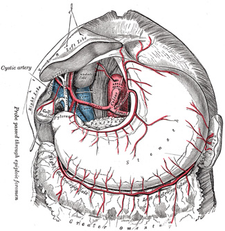 |
FIG. 532– The celiac artery and its branches; the liver has been raised, and the lesser omentum and anterior layer of the greater omentum removed. (See enlarged image) |
| |
| The right gastric artery (a. gastrica dextra; pyloric artery) arises from the hepatic, above the pylorus, descends to the pyloric end of the stomach, and passes from right to left along its lesser curvature, supplying it with branches, and anastomosing with the left gastric artery. | 11 |
| The gastroduodenal artery (a. gastroduodenalis) (Fig. 533) is a short but large branch, which descends, near the pylorus, between the superior part of the duodenum and the neck of the pancreas, and divides at the lower border of the duodenum into two branches, the right gastroepiploic and the superior pancreaticoduodenal. Previous to its division it gives off two or three small branches to the pyloric end of the stomach and to the pancreas. | 12 |
| The right gastroepiploic artery (a. gastroepiploica dextra) runs from right to left along the greater curvature of the stomach, between the layers of the greater omentum, anastomosing with the left gastroepiploic branch of the lienal artery. Except at the pylorus where it is in contact with the stomach, it lies about a finger's breadth from the greater curvature. This vessel gives off numerous branches, some of which ascend to supply both surfaces of the stomach, while others descend to supply the greater omentum and anastomose with branches of the middle colic. | 13 |
| The superior pancreaticoduodenal artery (a. pancreaticoduodenalis superior) descends between the contiguous margins of the duodenum and pancreas. It supplies both these organs, and anastomoses with the inferior pancreaticoduodenal branch of the superior mesenteric artery, and with the pancreatic branches of the lienal artery. | 14 |
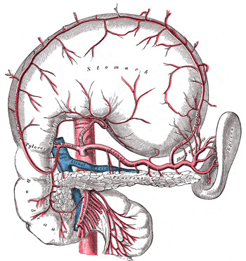 |
FIG. 533– The celiac artery and its branches; the stomach has been raised and the peritoneum removed. (See enlarged image) |
| |
| The cystic artery (a. cystica) (Fig. 532), usually a branch of the right hepatic, passes downward and forward along the neck of the gall-bladder, and divides into two branches, one of which ramifies on the free surface, the other on the attached surface of the gall-bladder. | 15 |
| 3. The Lienal or Splenic Artery (a. lienalis), the largest branch of the celiac artery, is remarkable for the tortuosity of its course. It passes horizontally to the left side, behind the stomach and the omental bursa of the peritoneum, and along the upper border of the pancreas, accompanied by the lienal vein, which lies below it; it crosses in front of the upper part of the left kidney, and, on arriving near the spleen, divides into branches, some of which enter the hilus of that organ between the two layers of the phrenicolienal ligament to be distributed to the tissues of the spleen; some are given to the pancreas, while others pass to the greater curvature of the stomach between the layers of the gastrolienal ligament. Its branches are: | 16 |
| Pancreatic. |
|
Short Gastric. |
|
Left Gastroepiploic. |
|
|
| The pancreatic branches (rami pancreatici) are numerous small vessels derived from the lienal as it runs behind the upper border of the pancreas, supplying its body and tail. One of these, larger than the rest, is sometimes given off near the tail of the pancreas; it runs from left to right near the posterior surface of the gland, following the course of the pancreatic duct, and is called the arteria pancreatica magna. These vessels anastomose with the pancreatic branches of the pancreaticoduodenal and superior mesenteric arteries. | 17 |
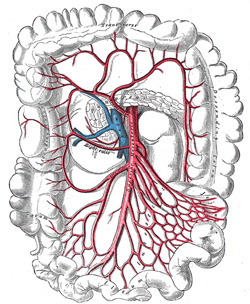 |
FIG. 534– The superior mesenteric artery and its branches. (See enlarged image) |
| |
| The short gastric arteries (aa. gastricæ breves; vasa brevia) consist of from five to seven small branches, which arise from the end of the lienal artery, and from its terminal divisions. They pass from left to right, between the layers of the gastrolienal ligament, and are distributed to the greater curvature of the stomach, anastomosing with branches of the left gastric and left gastroepiploic arteries. | 18 |
| The left gastroepiploic artery (a. gastroepiploica sinistra) the largest branch of the lienal, runs from left to right about a finger’s breadth or more from the greater curvature of the stomach, between the layers of the greater omentum, and anastomoses with the right gastroepiploic. In its course it distributes several ascending branches to both surfaces of the stomach; others descend to supply the greater omentum and anastomose with branches of the middle colic. | 19 |
| The superior mesenteric artery (a. mesenterica superior) (Fig. 534) is a large vessel which supplies the whole length of the small intestine, except the superior part of the duodenum; it also supplies the cecum and the ascending part of the colon and about one-half of the transverse part of the colon. It arises from the front of the aorta, about 1.25 cm. below the celiac artery, and is crossed at its origin by the lienal vein and the neck of the pancreas. It passes downward and forward, anterior to the processus uncinatus of the head of the pancreas and inferior part of the duodenum, and descends between the layers of the mesentery to the right iliac fossa, where, considerably diminished in size, it anastomoses with one of its own branches, viz., the ileocolic. In its course it crosses in front of the inferior vena cava, the right ureter and Psoas major, and forms an arch, the convexity of which is directed foward and downward to the left side, the concavity backward and upward to the right. It is accompanied by the superior mesenteric vein, which lies to its right side, and it is surrounded by the superior mesenteric plexus of nerves. | 20 |
| |
| Branches.—Its branches are: | 21 |
| Inferior Pancreaticoduodenal. |
|
Ileocolic. |
| Intestinal. |
|
Right Colic. |
| Middle Colic. |
|
| The Inferior Pancreaticoduodenal Artery (a. pancreaticoduodenalis inferior) is given off from the superior mesenteric or from its first intestinal branch, opposite the upper border of the inferior part of the duodenum. It courses to the right between the head of the pancreas and duodenum, and then ascends to anastomose with the superior pancreaticoduodenal artery. It distributes branches to the head of the pancreas and to the descending and inferior parts of the duodenum. | 22 |
| The Intestinal Arteries (aa. intestinales; vasa intestini tenuis) arise from the convex side of the superior mesenteric artery. They are usually from twelve to fifteen in number, and are distributed to the jejunum and ileum. They run nearly parallel with one another between the layers of the mesentery, each vessel dividing into two branches, which unite with adjacent branches, forming a series of arches, the convexities of which are directed toward the intestine (Fig. 535). From this first set of arches branches arise, which unite with similar branches from above and below and thus a second series of arches is formed; from the lower branches of the artery, a third, a fourth, or even a fifth series of arches may be formed, diminishing in size the nearer they approach the intestine. In the short, upper part of the mesentery only one set of arches exists, but as the depth of the mesentery increases, second, third, fourth, or even fifth groups are developed. From the terminal arches numerous small straight vessels arise which encircle the intestine, upon which they are distributed, ramifying between its coats. From the intestinal arteries small branches are given off to the lymph glands and other structures between the layers of the mesentery. | 23 |
| The Ileocolic Artery (a. ileocolica) is the lowest branch arising from the concavity of the superior mesenteric artery. It passes downward and to the right behind the peritoneum toward the right iliac fossa, where it divides into a superior and an inferior branch; the inferior anastomoses with the end of the superior mesenteric artery, the superior with the right colic artery. | 24 |
| The inferior branch of the ileocolic runs toward the upper border of the ileocolic junction and supplies the following branches (Fig. 536): | 25 |
| (a) colic, which pass upward on the ascending colon; (b) anterior and posterior cecal, which are distributed to the front and back of the cecum; (c) an appendicular artery, which descends behind the termination of the ileum and enters the mesenteriole of the vermiform process; it runs near the free margin of this mesenteriole and ends in branches which supply the vermiform process; and (d) ileal, which run upward and to the left on the lower part of the ileum, and anastomose with the termination of the superior mesenteric. | 26 |
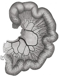 |
FIG. 535– Loop of small intestine showing distribution of intestinal arteries. (From a preparation by Mr. Hamilton Drummond.) The vessels were injected while the gut was in situ; the gut was then removed, and an x-ray photograph taken. (See enlarged image) |
| |
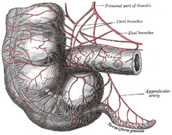 |
FIG. 536– Arteries of cecum and vermiform process. (See enlarged image) |
| |
| The Right Colic Artery (a. colica dextra) arises from about the middle of the concavity of the superior mesenteric artery, or from a stem common to it and the ileocolic. It passes to the right behind the peritoneum, and in front of the right internal spermatic or ovarian vessels, the right ureter and the Psoas major, toward the middle of the ascending colon; sometimes the vessel lies at a higher level, and crosses the descending part of the duodenum and the lower end of the right kidney. At the colon it divides into a descending branch, which anastomoses with the ileocolic, and an ascending branch, which anastomoses with the middle colic. These branches form arches, from the convexity of which vessels are distributed to the ascending colon. | 27 |
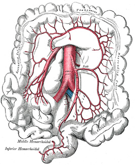 |
FIG. 537– The inferior mesenteric artery and its branches. (See enlarged image) |
| |
| The Middle Colic Artery (a. colica media) arises from the superior mesenteric just below the pancreas and, passing downward and forward between the layers of the transverse mesocolon, divides into two branches, right and left; the former anastomoses with the right colic; the latter with the left colic, a branch of the inferior mesenteric. The arches thus formed are placed about two fingers’ breadth from the transverse colon, to which they distribute branches. | 28 |
| The inferior mesenteric artery (a. mesenterica inferior) (Fig. 537) supplies the left half of the transverse part of the colon, the whole of the descending and iliac parts of the colon, the sigmoid colon, and the greater part of the rectum. It is smaller than the superior mesenteric, and arises from the aorta, about 3 or 4 cm. above its division into the common iliacs and close to the lower border of the inferior part of the duodenum. It passes downward posterior to the peritoneum, lying at first anterior to and then on the left side of the aorta. It crosses the left common iliac artery and is continued into the lesser pelvis under the name of the superior hemorrhoidal artery, which descends between the two layers of the sigmoid mesocolon and ends on the upper part of the rectum. | 29 |
| |
| Branches.—Its branches are: | 30 |
| Left Colic. |
|
Sigmoid. |
|
Superior Hemorrhoidal. |
|
|
| The Left Colic Artery (a. colica sinistra) runs to the left behind the peritoneum and in front of the Psoas major, and after a short, but variable, course divides into an ascending and a descending branch; the stem of the artery or its branches cross the left ureter and left internal spermatic vessels. The ascending branch crosses in front of the left kidney and ends, between the two layers of the transverse mesocolon, by anastomosing with the middle colic artery; the descending branch anastomoses with the highest sigmoid artery. From the arches formed by these anastomoses branches are distributed to the descending colon and the left part of the transverse colon. | 31 |
| The Sigmoid Arteries (aa. sigmoideæ) (Fig. 538), two or three in number, run obliquely downward and to the left behind the peritoneum and in front of the Psoas major, ureter, and internal spermatic vessels. Their branches supply the lower part of the descending colon, the iliac colon, and the sigmoid or pelvic colon; anastomosing above with the left colic, and below with the superior hemorrhoidal artery. | 32 |
| The Superior Hemorrhoidal Artery (a. hæmorrhoidalis superior) (Fig. 538), the continuation of the inferior mesenteric, descends into the pelvis between the layers of the mesentery of the sigmoid colon, crossing, in its course, the left common iliac vessels. It divides, opposite the third sacral vertebra, into two branches, which descend one on either side of the rectum, and about 10 or 12 cm. from the anus break up into several small branches. These pierce the muscular coat of the bowel and run downward, as straight vessels, placed at regular intervals from each other in the wall of the gut between its muscular and mucous coats, to the level of the Sphincter ani internus; here they form a series of loops around the lower end of the rectum, and communicate with the middle hemorrhoidal branches of the hypogastric, and with the inferior hemorrhoidal branches of the internal pudendal. | 33 |
| The middle suprarenal arteries (aa. suprarenales media; middle capsular arteries; suprarenal arteries) are two small vessels which arise, one from either side of the aorta, opposite the superior mesenteric artery. They pass lateralward and slightly upward, over the crura of the diaphragm, to the suprarenal glands, where they anastomose with suprarenal branches of the inferior phrenic and renal arteries. In the fetus these arteries are of large size. | 34 |
| The renal arteries (aa. renales) (Fig. 531), are two large trunks, which arise from the side of the aorta, immediately below the superior mesenteric artery. Each is directed across the crus of the diaphragm, so as to form nearly a right angle with the aorta. The right is longer than the left, on account of the position of the aorta; it passes behind the inferior vena cava, the right renal vein, the head of the pancreas, and the descending part of the duodenum. The left is somewhat higher than the right; it lies behind the left renal vein, the body of the pancreas and the lienal vein, and is crossed by the inferior mesenteric vein. Before reaching the hilus of the kidney, each artery divides into four or five branches; the greater number of these lie between the renal vein and ureter, the vein being in front, the ureter behind, but one or more branches are usually situated behind the ureter. Each vessel gives off some small inferior suprarenal branches to the suprarenal gland, the ureter, and the surrounding cellular tissue and muscles. One or two accessory renal arteries are frequently found, more especially on the left side they usually arise from the aorta, and may come off above or below the main artery, the former being the more common position. Instead of entering the kidney at the hilus, they usually pierce the upper or lower part of the gland. | 35 |
| The internal spermatic arteries (aa. spermaticæ internæ; spermatic arteries) (Fig. 531) are distributed to the testes. They are two slender vessels of considerable length, and arise from the front of the aorta a little below the renal arteries. Each passes obliquely downward and lateralward behind the peritoneum, resting on the Psoas major, the right spermatic lying in front of the inferior vena cava and behind the middle colic and ileocolic arteries and the terminal part of the ileum, the left behind the left colic and sigmoid arteries and the iliac colon. Each crosses obliquely over the ureter and the lower part of the external iliac artery to reach the abdominal inguinal ring, through which it passes, and accompanies the other constituents of the spermatic cord along the inguinal canal to the scrotum, where it becomes tortuous, and divides into several branches. Two or three of these accompany the ductus deferens, and supply the epididymis, anastomosing with the artery of the ductus deferens; others pierce the back part of the tunica albuginea, and supply the substance of the testis. The internal spermatic artery supplies one or two small branches to the ureter, and in the inguinal canal gives one or two twigs to the Cremaster. | 36 |
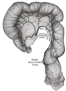 |
FIG. 538– Sigmoid colon and rectum, showing distribution of branches of inferior mesenteric artery and their anastomoses. (From a preparation by Mr. Hamilton Drummond.) Prepared in same manner as Fig. 535. (See enlarged image) |
| |
| The ovarian arteries (aa. ovaricæ) are the corresponding arteries in the female to the internal spermatic in the male. They supply the ovaries, are shorter than the internal spermatics, and do not pass out of the abdominal cavity. The origin and course of the first part of each artery are the same as those of the internal spermatic, but on arriving at the upper opening of the lesser pelvis the ovarian artery passes inward, between the two layers of the ovariopelvic ligament and of the broad ligament of the uterus, to be distributed to the ovary. Small branches are given to the ureter and the uterine tube, and one passes on to the side of the uterus, and unites with the uterine artery. Other offsets are continued on the round ligament of the uterus, through the inguinal canal, to the integument of the labium majus and groin. | 37 |
| At an early period of fetal life, when the testes or ovaries lie by the side of the vertebral column, below the kidneys, the internal spermatic or ovarian arteries are short; but with the descent of these organs into the scrotum or lesser pelvis, the arteries are gradually lengthened. | 38 |
| The inferior phrenic arteries (aa. phrenicæ inferiores) (Fig. 531) are two small vessels, which supply the diaphragm but present much variety in their origin. They may arise separately from the front of the aorta, immediately above the celiac artery, or by a common trunk, which may spring either from the aorta or from the celiac artery. Sometimes one is derived from the aorta, and the other from one of the renal arteries; they rarely arise as separate vessels from the aorta. They diverge from one another across the crura of the diaphragm, and then run obliquely upward and lateralward upon its under surface. The left phrenic passes behind the esophagus, and runs forward on the left side of the esophageal hiatus. The right phrenic passes behind the inferior vena cava, and along the right side of the foramen which transmits that vein. Near the back part of the central tendon each vessel divides into a medial and a lateral branch. The medial branch curves forward, and anastomoses with its fellow of the opposite side, and with the musculophrenic and pericardiacophrenic arteries. The lateral branch passes toward the side of the thorax, and anastomoses with the lower intercostal arteries, and with the musculophrenic. The lateral branch of the right phrenic gives off a few vessels to the inferior vena cava; and the left one, some branches to the esophagus. Each vessel gives off superior suprarenal branches to the suprarenal gland of its own side. The spleen and the liver also receive a few twigs from the left and right vessels respectively. | 39 |
| The lumbar arteries (aa. lumbales) are in series with the intercostals. They are usually four in number on either side, and arise from the back of the aorta, opposite the bodies of the upper four lumbar vertebræ. A fifth pair, small in size, is occasionally present: they arise from the middle sacral artery. They run lateralward and backward on the bodies of the lumbar vertebræ, behind the sympathetic trunk, to the intervals between the adjacent transverse processes, and are then continued into the abdominal wall. The arteries of the right side pass behind the inferior vena cava, and the upper two on each side run behind the corresponding crus of the diaphragm. The arteries of both sides pass beneath the tendinous arches which give origin to the Psoas major, and are then continued behind this muscle and the lumbar plexus. They now cross the Quadratus lumborum, the upper three arteries running behind, the last usually in front of the muscle. At the lateral border of the Quadratus lumborum they pierce the posterior aponeurosis of the Transversus abdominis and are carried forward between this muscle and the Obliquus internus. They anastomose with the lower intercostal, the subcostal, the iliolumbar, the deep iliac circumflex, and the inferior epigastric arteries. | 40 |
| |
| Branches.—In the interval between the adjacent transverse processes each lumbar artery gives off a posterior ramus which is continued backward between the transverse processes and is distributed to the muscles and skin of the back; it furnishes a spinal branch which enters the vertebral canal and is distributed in a manner similar to the spinal branches of the posterior rami of the intercostal arteries (page 601). Muscular branches are supplied from each lumbar artery and from its posterior ramus to the neighboring muscles. | 41 |
| The middle sacral artery (a. sacralis media) (Fig. 531) is a small vessel, which arises from the back of the aorta, a little above its bifurcation. It descends in the middle line in front of the fourth and fifth lumbar vertebræ, the sacrum and coccyx, and ends in the glomus coccygeum (coccygeal gland). From it, minute branches are said to pass to the posterior surface of the rectum. On the last lumbar vertebra it anastomoses with the lumbar branch of the iliolumbar artery; in front of the sacrum it anastomoses with the lateral sacral arteries, and sends offsets into the anterior sacral foramina. It is crossed by the left common iliac vein, and is accompanied by a pair of venæ comitantes; these unite to form a single vessel, which opens into the left common iliac vein. | 42 |
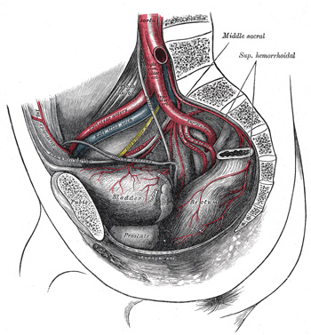 |
FIG. 539– The arteries of the pelvis. (See enlarged image) |
| |


