(N. Opticus; Second Nerve)
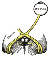
The optic nerve (Fig. 773), or nerve of sight, consists mainly of fibers derived from the ganglionic cells of the retina. These axons terminate in arborizations around the cells in the lateral geniculate body, pulvinar, and superior colliculus which constitute the lower or primary visual centers. From the cells of the lateral geniculate body and the pulvinar fibers pass to the cortical visual center, situated in the cuneus and in the neighborhood of the calcarine fissure. A few fibers of the optic nerve, of small caliber, pass from the primary centers to the retina and are supposed to govern chemical changes in the retina and also the movements of some of its elements (pigment cells and cones). There are also a few fine fibers, afferent fibers, extending from the retina to the brain, that are supposed to be concerned in pupillary reflexes. | 1 |
 |
FIG. 773– The left optic nerve and the optic tracts. (See enlarged image) |
| |
| The optic nerve is peculiar in that its fibers and ganglion cells are probably third in the series of neurons from the receptors to the brain. Consequently the optic nerve corresponds rather to a tract of fibers within the brain than to the other cranial nerves. Its fibers pass backward and medialward through the orbit and optic foramen to the optic commissure where they partially decussate. The mixed fibers from the two nerves are continued in the optic tracts, the primary visual centers of the brain. | 2 |
| The orbital portion of the optic nerve is from 20 mm. to 30 mm. in length and has a slightly sinuous course to allow for movements of the eyeball. It is invested by an outer sheath of dura mater and an inner sheath from the arachnoid which are attached to the sclera around the area where the nerve fibers pierce the choroid and sclera of the bulb. A little behind the bulb of the eye the central artery of the retina with its accompanying vein perforates the optic nerve, and runs within it to the retina. As the nerve enters the optic foramen its dural sheath becomes continuous with that lining the orbit and the optic foramen. In the optic foramen the ophthalmic artery lies below and to its outer side. The intercranial portion of the optic nerve is about 10 mm. in length. | 3 |
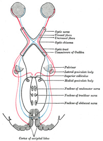
| The Optic Chiasma (chiasma opticum), somewhat quadrilateral in form, rests upon the tuberculum sellæ and on the anterior part of the diaphragma sellæ. It is in relation, above, with the lamina terminalis; behind, with the tuber cinereum; on either side, with the anterior perforated substance. Within the chiasma, the optic nerves undergo a partial decussation. The fibers forming the medial part of each tract and posterior part of the chiasma have no connection with the optic nerves. They simply cross in the chiasma, and connect the medial geniculate bodies of the two sides; they form the commissure of Gudden. The remaining and principal part of the chiasma consists of two sets of fibers, crossed and uncrossed. The crossed fibers which are the more numerous, occupy the central part of the chiasma, and pass from the optic nerve of one side to the optic tract of the other, decussating in the chiasma with similar fibers of the opposite optic nerve. The uncrossed fibers occupy the lateral part of the chiasma, and pass from the nerve of one side into the tract of the same side. 130 | 4 |
 |
FIG. 774– Scheme showing central connections of the optic nerves and optic tracts. (See enlarged image) |
| |
| The crossed fibers of the optic nerve tend to occupy the medial side of the nerve and the uncrossed fibers the lateral side. In the optic tract, however, the fibers are much more intermingled. | 5 |
| The Optic Tract (Fig. 774), passes backward and outward from the optic chiasma over the tuber cinereum and anterior perforated space to the cerebral peduncle and winds obliquely across its under surface. Its fibers terminate in the lateral geniculate body, the pulvinar and the superior colliculus. It is adherent to the tuber cinereum and the cerebral peduncle as it passes over them. In the region of the lateral geniculate body it splits into two bands. The medial and smaller one is a part of the commissure of Gudden and ends in the medial geniculate body. | 6 |
| From its mode of development, and from its structure, the optic nerve must be regarded as a prolongation of the brain substance, rather than as an ordinary cerebrospinal nerve. As it passes from the brain it receives sheaths from the three cerebral membranes, a perineural sheath from the pia mater, an intermediate sheath from the arachnoid, and an outer sheath from the dura mater, which is also connected with the periosteum as it passes through the optic foramen. These sheaths are separated from each other by cavities which communicate with the subdural and subarachnoid cavities respectively. The innermost or perineural sheath sends a process around the arteria centralis retinæ into the interior of the nerve, and enters intimately into its structure. | 7 |

