(Cavum Oris; Oral Or Buccal Cavity)
The cavity of the mouth is placed at the commencement of the digestive tube (Fig. 994); it is a nearly oval-shaped cavity which consists of two parts: an outer, smaller portion, the vestibule, and an inner, larger part, the mouth cavity proper. | 1 |
| The Vestibule (vestibulum oris) is a slit-like space, bounded externally by the lips and cheeks; internally by the gums and teeth. It communicates with the surface of the body by the rima or orifice of the mouth. Above and below, it is limited by the reflection of the mucous membrane from the lips and cheeks to the gum covering the upper and lower alveolar arch respectively. It receives the secretion from the parotid salivary glands, and communicates, when the jaws are closed, with the mouth cavity proper by an aperture on either side behind the wisdom teeth, and by narrow clefts between opposing teeth. | 2 |
| The Mouth Cavity Proper (cavum oris proprium) (Fig. 1014) is bounded laterally and in front by the alveolar arches with their contained teeth; behind, it communicates with the pharynx by a constricted aperture termed the isthmus faucium. It is roofed in by the hard and soft palates, while the greater part of the floor is formed by the tongue, the remainder by the reflection of the mucous membrane from the sides and under surface of the tongue to the gum lining the inner aspect of the mandible. It receives the secretion from the submaxillary and sublingual salivary glands. | 3 |
| |
| Structure.—The mucous membrane lining the mouth is continuous with the integument at the free margin of the lips, and with the mucous lining of the pharynx behind; it is of a rosepink tinge during life, and very thick where it overlies the hard parts bounding the cavity. It is covered by stratified squamous epithelium. | 4 |
| The Lips (labia oris), the two fleshy folds which surround the rima or orifice of the mouth, are formed externally of integument and internally of mucous membrane, between which are found the Orbicularis oris muscle, the labial vessels, some nerves, areolar tissue, and fat, and numerous small labial glands. The inner surface of each lip is connected in the middle line to the corresponding gum by a fold of mucous membrane, the frenulum—the upper being the larger. | 5 |
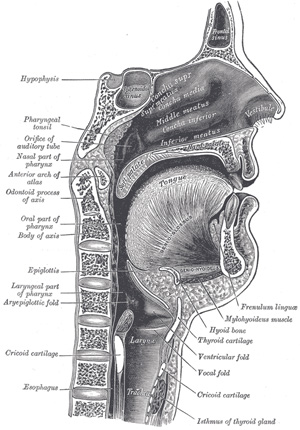 |
FIG. 994– Sagittal section of nose mouth, pharynx, and larynx. (See enlarged image) |
| |
| The Labial Glands (glandulœ labiales) are situated between the mucous membrane and the Orbicularis oris, around the orifice of the mouth. They are circular in form, and about the size of small peas; their ducts open by minute orifices upon the mucous membrane. In structure they resemble the salivary glands. | 6 |
| The Cheeks (buccæ) form the sides of the face, and are continuous in front with the lips. They are composed externally of integument; internally of mucous membrane; and between the two of a muscular stratum, besides a large quantity of fat, areolar tissue, vessels, nerves, and buccal glands. | 7 |
| |
| Structure.—The mucous membrane lining the cheek is reflected above and below upon the gums, and is continuous behind with the lining membrane of the soft palate. Opposite the second molar tooth of the maxilla is a papilla, on the summit of which is the aperture of the parotid duct. The principal muscle of the cheek is the Buccinator; but other muscles enter into its formation, viz., the Zygomaticus, Risorius, and Platysma. | 8 |
| The buccal glands are placed between the mucous membrane and Buccinator muscle: they are similar in structure to the labial glands, but smaller. About five, of a larger size than the rest, are placed between the Masseter and Buccinator muscles around the distal extremity of the parotid duct; their ducts open in the mouth opposite the last molar tooth. They are called molar glands. | 9 |
| The Gums (gingivœ) are composed of dense fibrous tissue, closely connected to the periosteum of the alveolar processes, and surrounding the necks of the teeth. They are covered by smooth and vascular mucous membrane, which is remarkable for its limited sensibility. Around the necks of the teeth this membrane presents numerous fine papillæ, and is reflected into the alveoli, where it is continuous with the periosteal membrane lining these cavities. | 10 |
| The Palate (palatum) forms the roof of the mouth; it consists of two portions, the hard palate in front, the soft palate behind. | 11 |
| The Hard Palate (palatum durum) (Fig. 1014) is bounded in front and at the sides by the alveolar arches and gums; behind, it is continuous with the soft palate. It is covered by a dense structure, formed by the periosteum and mucous membrane of the mouth, which are intimately adherent. Along the middle line is a linear raphæ, which ends anteriorly in a small papilla corresponding with the incisive canal. On either side and in front of the raphé the mucous membrane is thick, pale in color, and corrugated; behind, it is thin, smooth, and of a deeper color; it is covered with stratified squamous epithelium, and furnished with numerous palatal glands, which lie between the mucous membrane and the surface of the bone. | 12 |
| The Soft Palate (palatum molle) (Fig. 1014) is a movable fold, suspended from the posterior border of the hard palate, and forming an incomplete septum between the mouth and pharynx. It consists of a fold of mucous membrane enclosing muscular fibers, an aponeurosis, vessels, nerves, adenoid tissue, and mucous glands. When occupying its usual position, i. e., relaxed and pendent, its anterior surface is concave, continuous with the roof of the mouth, and marked by a median raphé. Its posterior surface is convex, and continuous with the mucous membrane covering the floor of the nasal cavities. Its upper border is attached to the posterior margin of the hard palate, and its sides are blended with the pharynx. Its lower border is free. Its lower portion, which hangs like a curtain between the mouth and pharynx is termed the palatine velum. | 13 |
| Hanging from the middle of its lower border is a small, conical, pendulous process, the palatine uvula; and arching lateralward and downward from the base of the uvula on either side are two curved folds of mucous membrane, containing muscular fibers, called the arches or pillars of the fauces. | 14 |
| |
| The Teeth (dentes) (Figs. 995 to 997).—Man is provided with two sets of teeth, which make their appearance at different periods of life. Those of the first set appear in childhood, and are called the deciduous or milk teeth. Those of the second set, which also appear at an early period, may continue until old age, and are named permanent. | 15 |
| The deciduous teeth are twenty in number: four incisors, two canines, and four molars, in each jaw. | 16 |
| The permanent teeth are thirty-two in number: four incisors, two canines, four premolars, and six molars, in each jaw. | 17 |
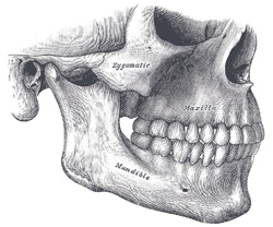 |
FIG. 995– Side view of the teeth and jaws. (See enlarged image) |
| |
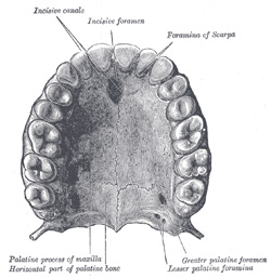 |
FIG. 996– Permanent teeth of upper dental arch, seen from below. (See enlarged image) |
| |
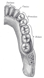 |
FIG. 997– Permanent teeth of right half of lower dental arch, seen from above. (See enlarged image) |
| |
| The dental formulæ may be represented as follows: | 18 |
| Deciduous Teeth. |
|
|
mol. | can. | in. | in. | can. | mol. |
|
|
| Upper jaw |
| 2 | 1 | 2 | 2 | 1 | 2 |
| Total 20 |
| Lower jaw |
| 2 | 1 | 2 | 2 | 1 | 2 |
|
| Permanent Teeth. |
|
mol. | pr.mol. | can. | in. | in. | can. | pr.mol. | mol. |
|
| Upper jaw | 3 | 2 | 1 | 2 | 2 | 1 | 2 | 3 | Total 32 |
| Lower jaw | 3 | 2 | 1 | 2 | 2 | 1 | 2 | 3 |
|
| |
| General Characteristics.—Each tooth consists of three portions: the crown, projecting above the gum; the root, imbedded in the alveolus; and the neck, the constricted portion between the crown and root. | 19 |
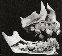 |
FIG. 998– Maxillæ at about one year. (Noyes.) (See enlarged image) |
| |
| The roots of the teeth are firmly implanted in depressions within the alveoli; these depressions are lined with periosteum which invests the tooth as far as the neck. At the margins of the alveoli, the periosteum is continuous with the fibrous structure of the gums. | 20 |
| In consequence of the curve of the dental arch, terms such as anterior and posterior, as applied to the teeth, are misleading and confusing. Special terms are therefore used to indicate the different surfaces of a tooth: the surface directed toward the lips or cheek is known as the labial or buccal surface; that directed toward the tongue is described as the lingual surface; those surfaces which touch neighboring teeth are termed surfaces of contact. In the case of the incisor and canine teeth the surfaces of contact are medial and lateral; in the premolar and molar teeth they are anterior and posterior. | 21 |
| The superior dental arch is larger than the inferior, so that in the normal condition the teeth in the maxillæ slightly overlap those of the mandible both in front and at the sides. Since the upper central incisors are wider than the lower, the other teeth in the upper arch are thrown somewhat distally, and the two sets do not quite correspond to each other when the mouth is closed: thus the upper canine tooth rests partly on the lower canine and partly on the first premolar, and the cusps of the upper molar teeth lie behind the corresponding cusps of the lower molar teeth. The two series, however, end at nearly the same point behind; this is mainly because the molars in the upper arch are the smaller. | 22 |
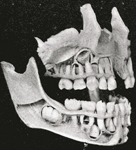 |
FIG. 999– The complete temporary dentition (about three years), showing the relation of the developing permanent teeth. (Noyes.) (See enlarged image) |
| |
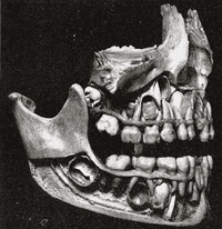 |
FIG. 1000– The complete temporary dentition and the first permanent molar. Note the relation of the bicuspids to the temporary molars. (In the seventh year.) (Noyes.) (See enlarged image) |
| |
| |
| The Permanent Teeth (dentes permanentes) (Figs. 1002, 1003).—The Incisors (dentes incisivi; incisive or cutting teeth) are so named from their presenting a sharp cutting edge, adapted for biting the food. They are eight in number, and form the four front teeth in each dental arch. | 23 |
| The crown is directed vertically, and is chisel-shaped, being bevelled at the expense of its lingual surface, so as to present a sharp horizontal cutting edge, which, before being subjected to attrition, presents three small prominent points separated by two slight notches. It is convex, smooth, and highly polished on its labial surface; concave on its lingual surface, where, in the teeth of the upper arch, it is frequently marked by an inverted V-shaped eminence, situated near the gum. This is known as the basal ridge or cingulum. The neck is constricted. The root is long, single, conical, transversely flattened, thicker in front than behind, and slightly grooved on either side in the longitudinal direction. | 24 |
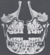 |
FIG. 1001– Front view of the skull shown in Fig. 1000. Note the relation of the permanent incisors and cuspids to each other and the roots of the temporary teeth. (Noyes.) (See enlarged image) |
| |
| The upper incisors are larger and stronger than the lower, and are directed obliquely downward and forward. The central ones are larger than the lateral, and their roots are more rounded. | 25 |
| The lower incisors are smaller than the upper: the central ones are smaller than the lateral, and are the smallest of all the incisors. They are placed vertically and are somewhat bevelled in front, where they have been worn down by contact with the overlapping edge of the upper teeth. The cingulum is absent. | 26 |
| The Canine Teeth (dentes canini) are four in number, two in the upper, and two in the lower arch, one being placed laterally to each lateral incisor. They are larger and stronger than the incisors, and their roots sink deeply into the bones, and cause well-marked prominences upon the surface. | 27 |
| The crown is large and conical, very convex on its labial surface, a little hollowed and uneven on its lingual surface, and tapering to a blunted point or cusp, which projects beyond the level of the other teeth. The root is single, but longer and thicker than that of the incisors, conical in form, compressed laterally, and marked by a slight groove on each side. | 28 |
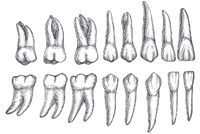 |
FIG. 1002– Permanent teeth. Right side. (Burchard.) (See enlarged image) |
| |
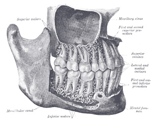 |
FIG. 1003– The permanent teeth, viewed from the right. The external layer of bone has been partly removed and the maxillary sinus has been opened. (Spalteholz.) (See enlarged image) |
| |
| The upper canine teeth (popularly called eye teeth) are larger and longer than the lower, and usually present a distinct basal ridge. | 29 |
| The lower canine teeth (popularly called stomach teeth) are placed nearer the middle line than the upper, so that their summits correspond to the intervals between the upper canines and the lateral incisors. | 30 |
| The Premolars or Bicuspid teeth (dentes præmolares) are eight in number, four in each arch. They are situated lateral to and behind the canine teeth, and are smaller and shorter than they. | 31 |
| The crown is compressed antero-posteriorly, and surmounted by two pyramidal eminences or cusps, a labial and a lingual, separated by a groove; hence their name bicuspid. Of the two cusps the labial is the larger and more prominent. The neck is oval. The root is generally single, compressed, and presents in front and behind a deep groove, which indicates a tendency in the root to become double. The apex is generally bifid. | 32 |
| The upper premolars are larger, and present a greater tendency to the division of their roots than the lower; this is especially the case in the first upper premolar. | 33 |
| The Molar Teeth (dentes molares) are the largest of the permanent set, and their broad crowns are adapted for grinding and pounding the food. They are twelve in number; six in each arch, three being placed posterior to each of the second premolars. | 34 |
| The crown of each is nearly cubical in form, convex on its buccal and lingual surfaces, flattened on its surfaces of contact; it is surmounted by four or five tubercles, or cusps, separated from each other by a crucial depression; hence the molars are sometimes termed multicuspids. The neck is distinct, large, and rounded. | 35 |
| Upper Molars.—As a rule the first is the largest, and the third the smallest of the upper molars. The crown of the first has usually four tubercles; that of the second, three or four; that of the third, three. Each upper molar has three roots, and of these two are buccal and nearly parallel to one another; the third is lingual and diverges from the others as it runs upward. The roots of the third molar (dens serotinus or wisdom-tooth) are more or less fused together. | 36 |
| |
| Lower Molars.—The lower molars are larger than the upper. On the crown of the first there are usually five tubercles; on those of the second and third, four or five. Each lower molar has two roots, an anterior, nearly vertical, and a posterior, directed obliquely backward; both roots are grooved longitudinally, indicating a tendency to division. The two roots of the third molar (dens serotinus or wisdom tooth) are more or less united. | 37 |
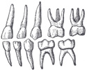 |
FIG. 1004– Deciduous teeth. Left side. (See enlarged image) |
| |
| |
| The Deciduous Teeth (dentes decidui; temporary or milk teeth) (Fig. 1004).—The deciduous are smaller than, but, generally speaking, resemble in form, the teeth which bear the same names in the permanent set. The hinder of the two molars is the largest of all the deciduous teeth, and is succeeded by the second premolar. The first upper molar has only three cusps—two labial, one lingual; the second upper molar has four cusps. The first lower molar has four cusps; the second lower molar has five. The roots of the deciduous molars are smaller and more divergent than those of the permanent molars, but in other respects bear a strong resemblance to them. | 38 |
| |
| Structure of the Teeth.—On making a vertical section of a tooth (Fig. 1005), a cavity will be found in the interior of the crown and the center of each root; it opens by a minute orifice at the extremity of the latter. This is called the pulp cavity, and contains the dental pulp, a loose connective tissue richly supplied with vessels and nerves, which enter the cavity through the small aperture at the point of each root. Some of the cells of the pulp are arranged as a layer on the wall of the pulp cavity; they are named the odontoblasts of Waldeyer, and during the development of the tooth, are columnar in shape, but later on, after the dentin is fully formed, they become flattened and resemble osteoblasts. Each has two fine processes, the outer one passing into a dental canaliculus, the inner being continuous with the processes of the connective-tissue cells of the pulp matrix. | 39 |
| The solid portion of the tooth consists of (1) the ivory or dentin, which forms the bulk of the tooth; (2) the enamel, which covers the exposed part of the crown; and (3) a thin layer of bone, the cement or crusta petrosa, which is disposed on the surface of the root. | 40 |
| The dentin (substantia eburnea; ivory) (Fig. 1007) forms the principal mass of a tooth. It is a modification of osseous tissue, from which it differs, however, in structure. On microscopic examination it is seen to consist of a number of minute wavy and branching tubes, the dental canaliculi, imbedded in a dense homogeneous substance, the matrix. | 41 |
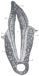 |
FIG. 1005– Vertical section of a tooth in situ. X 15. c is placed in the pulp cavity, opposite the neck of the tooth; the part above it is the crown, that below is the root. 1. Enamel with radial and concentric markings. 2. Dentin with tubules and incremental lines. 3. Cement or crusta petrosa, with bone corpuscles. 4. Dental periosteum. 5. Mandible. (See enlarged image) |
| |
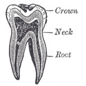 |
FIG. 1006– Vertical section of a molar tooth. (See enlarged image) |
| |
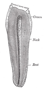 |
FIG. 1007– Vertical section of a premolar tooth. (Magnified.) (See enlarged image) |
| |
| The dental canaliculi (dentinal tubules) (Fig. 1008) are placed parallel with one another, and open at their inner ends into the pulp cavity. In their course to the periphery they present two or three curves, and are twisted on themselves in a spiral direction. These canaliculi vary in direction: thus in a tooth of the mandible they are vertical in the upper portion of the crown, becoming oblique and then horizontal in the neck and upper part of the root, while toward the lower part of the root they are inclined downward. In their course they divide and subdivide dichotomously, and, especially in the root, give off minute branches, which join together in loops in the matrix, or end blindly. Near the periphery of the dentin, the finer ramifications of the canaliculi terminate imperceptibly by free ends. The dental canaliculi have definite walls, consisting of an elastic homogeneous membrane, the dentinal sheath of Neumann, which resists the action of acids; they contain slender cylindrical prolongations of the odontoblasts, first described by Tomes, and named Tomes’ fibers or dentinal fibers. | 42 |
| The matrix (intertubular dentin) is translucent, and contains the chief part of the earthy matter of the dentin. In it are a number of fine fibrils, which are continuous with the fibrils of the dental pulp. After the earthy matter has been removed by steeping a tooth in weak acid, the animal basis remaining may be torn into laminæ which run parallel with the pulp cavity, across the direction of the tubes. A section of dry dentin often displays a series of somewhat parallel lines—the incremental lines of Salter. These lines are composed of imperfectly calcified dentin arranged in layers. In consequence of the imperfection in the calcifying process, little irregular cavities are left, termed interglobular spaces (Fig. 1008). Normally a series of these spaces is found toward the outer surface of the dentin, where they form a layer which is sometimes known as the granular layer. They have received their name from the fact that they are surrounded by minute nodules or globules of dentin. Other curved lines may be seen parallel to the surface. These are the lines of Schreger, and are due to the optical effect of simultaneous curvature of the dentinal fibers. | 43 |
| Chemical Composition.—According to Berzelius and von Bibra, dentin consists of 28 parts of animal and 72 parts of earthy matter. The animal matter is converted by boiling into gelatin. The earthy matter consists of phosphate of lime, carbonate of lime, a trace of fluoride of calcium, phosphate of magnesium, and other salts. | 44 |
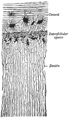 |
FIG. 1008– Transverse section of a portion of the root of a canine tooth. X 300. (See enlarged image) |
| |
| The enamel (substantia adamantina) is the hardest and most compact part of the tooth, and forms a thin crust over the exposed part of the crown, as far as the commencement of the root. It is thickest on the grinding surface of the crown, until worn away by attrition, and becomes thinner toward the neck. It consists of minute hexagonal rods or columns termed enamel fibers or enamel prisms (prismata adamantina). They lie parallel with one another, resting by one extremity upon the dentin, which presents a number of minute depressions for their reception; and forming the free surface of the crown by the other extremity. The columns are directed vertically on the summit of the crown, horizontally at the sides; they are about 4μ in diameter, and pursue a more or less wavy course. Each column is a six-sided prism and presents numerous dark transverse shadings; these shadings are probably due to the manner in which the columns are developed in successive stages, producing shallow constrictions, as will be subsequently explained. Another series of lines, having a brown appearance, the parallel striæ or colored lines of Retzius, is seen on section. According to Ebner, they are produced by air in the interprismatic spaces; others believe that they are the result of true pigmentation. | 45 |
| Numerous minute interstices intervene between the enamel fibers near their dentinal ends, a provision calculated to allow of the permeation of fluids from the dental canaliculi into the substance of the enamel. | 46 |
| Chemical Composition.—According to von Bibra, enamel consists of 96.5 per cent. of earthy matter, and 3.5 per cent. of animal matter. The earthy matter consists of phosphate of lime, with traces of fluoride of calcium, carbonate of lime, phosphate of magnesium, and other salts. According to Tomes, the enamel contains the merest trace of organic matter. | 47 |
| The crusta petrosa or cement (substantia ossea) is disposed as a thin layer on the roots of the teeth, from the termination of the enamel to the apex of each root, where it is usually very thick. In structure and chemical composition it resembles bone. It contains, sparingly, the lacunæ and canaliculi which characterize true bone; the lacunæ placed near the surface receive the canaliculi radiating from the side of the lacunæ toward the periodontal membrane; and those more deeply placed join with the adjacent dental canaliculi. In the thicker portions of the crusta petrosa, the lamellæ and Haversian canals peculiar to bone are also found. | 48 |
| As age advances, the cement increases in thickness, and gives rise to those bony growths or exostoses so common in the teeth of the aged; the pulp cavity also becomes partially filled up by a hard substance, intermediate in structure between dentin and bone (osteodentin, Owen; secondary dentin, Tomes). It appears to be formed by a slow conversion of the dental pulp, which shrinks, or even disappears. | 49 |
| |
| Development of the Teeth (Figs. 1009 to 1012).—In describing the development of the teeth, the mode of formation of the deciduous teeth must first be considered, and then that of the permanent series. | 50 |
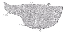 |
FIG. 1009– Sagittal section through the first lower deciduous molar of a human embryo 30 mm. long. (Röse.) X 100. L.E.L. Labiodental lamina, here separated from the dental lamina. Z.L. Placed over the shallow dental furrow, points to the dental lamina, which is spread out below to form the enamel germ of the future tooth. P.p. Bicuspidate papilla, capped by the enamel germ. Z.S. Condensed tissue forming dental sac. M.E. Mouth epithelium (See enlarged image) |
| |
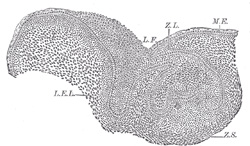 |
FIG. 1010– Similar section through the canine tooth of an embryo 40 mm. long. (Röse.) X 100. L.F. Labio dental furrow. The other lettering as in Fig. 1009. (See enlarged image) |
| |
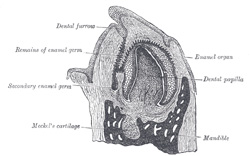 |
FIG. 1011– Vertical section of the mandible of an early human fetus. X 25. (See enlarged image) |
| |
| |
| Development of the Deciduous Teeth.—The development of the deciduous teeth begins about the sixth week of fetal life as a thickening of the epithelium along the line of the future jaw, the thickening being due to a rapid multiplication of the more deeply situated epithelial cells. As the cells multiply they extend into the subjacent mesoderm, and thus form a ridge or strand of cells imbedded in mesoderm. About the seventh week a longitudinal splitting or cleavage of this strand of cells takes place, and it becomes divided into two strands; the separation begins in front and extends laterally, the process occupying four or five weeks. Of the two strands thus formed, the labial forms the labiodental lamina; while the other, the lingual, is the ridge of cells in connection with which the teeth, both deciduous and permanent, are developed. Hence it is known as the dental lamina or common dental germ. It forms a flat band of cells, which grows into the substance of the embryonic jaw, at first horizontally inward, and then, as the teeth develop, vertically, i. e., upward in the upper jaw, and downward in the lower jaw. While still maintaining a horizontal direction it has two edges—an attached edge, continuous with the epithelium lining the mouth, and a free edge, projecting inward, and imbedded in the mesodermal tissue of the embryonic jaw. Along its line of attachment to the buccal epithelium is a shallow groove, the dental furrow. | 51 |
| About the ninth week the dental lamina begins to develop enlargements along its free border. These are ten in number in each jaw, and each corresponds to a future deciduous tooth. They consist of masses of epithelial cells; and the cells of the deeper part—that is, the part farthest from the margin of the jaw—increase rapidly and spread out in all directions. Each mass thus comes to assume a club shape, connected with the general epithelial lining of the mouth by a narrow neck, embraced by mesoderm. They are now known as special dental germs. After a time the lower expanded portion inclines outward, so as to form an angle with the superficial constricted portion, which is sometimes known as the neck of the special dental germ. About the tenth week the mesodermal tissue beneath these special dental germs becomes differentiated into papillæ; these grow upward, and come in contact with the epithelial cells of the special dental germs, which become folded over them like a hood or cap. There is, then, at this stage a papilla (or papillæ) which has already begun to assume somewhat the shape of the crown of the future tooth, and from which the dentin and pulp of the tooth are formed, surmounted by a dome or cap of epithelial cells from which the enamel is derived. | 52 |
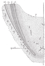 |
FIG. 1012– Longitudinal section of the lower part of a growing tooth, showing the extension of the layer of adamantoblasts beyond the crown to mark off the limit of formation of the dentin of the root. (Röse.) ad. Adamantoblasts, continuous below with ep.sch., the epithelial sheath of Hertwig. d. Dentin. en. Enamel. od. Odontoblasts. p. Pulp. (See enlarged image) |
| |
| In the meantime, while these changes have been going on, the dental lamina has been extending backward behind the special dental germ corresponding to the second deciduous molar tooth, and at about the seventeenth week it presents an enlargement, the special dental germ, for the first permanent molar, soon followed by the formation of a papilla in the mesodermal tissue for the same tooth. This is followed, about the sixth month after birth, by a further extension backward of the dental lamina, with the formation of another enlargement and its corresponding papilla for the second molar. And finally the process is repeated for the third molar, its papilla appearing about the fifth year of life. | 53 |
| After the formation of the special dental germs, the dental lamina undergoes atrophic changes and becomes cribriform, except on the lingual and lateral aspects of each of the special germs of the temporary teeth, where it undergoes a local thickening forming the special dental germ of each of the successional permanent teeth—i. e., the ten anterior ones in each jaw. Here the same process goes on as has been described in connection with those of the deciduous teeth: that is, they recede into the substance of the gum behind the germs of the deciduous teeth. As they recede they become club-shaped, form expansions at their distal extremities, and finally meet papillæ, which have been formed in the mesoderm, just in the same manner as was the case in the deciduous teeth. The apex of each papilla indents the dental germ, which encloses it, and, forming a cap for it, becomes converted into the enamel, while the papilla forms the dentin and pulp of the permanent tooth. | 54 |
| The special dental germs consist at first of rounded or polyhedral epithelial cells; after the formation of the papillæ, these cells undergo a differentiation into three layers. Those which are in immediate contact with the papilla become elongated, and form a layer of well-marked columnar epithelium coating the papilla. They are the cells which form the enamel fibers, and are therefore termed enamel cells or adamantoblasts. The cells of the outer layer of the special dental germ, which are in contact with the inner surface of the dental sac, presently to be described, are much shorter, cubical in form, and are named the external enamel epithelium. All the intermediate round cells of the dental germ between these two layers undergo a peculiar change. They become stellate in shape and develop processes, which unite to form a net-work into which fluid is secreted; this has the appearance of a jelly, and to it the name of enamel pulp is given. This transformed special dental germ is now known under the name of enamel organ (Fig. 1011). | 55 |
| While these changes are going on, a sac is formed around each enamel organ from the surrounding mesodermal tissue. This is known as the dental sac, and is a vascular membrane of connective tissue. It grows up from below, and thus encloses the whole tooth germ; as it grows it causes the neck of the enamel organ to atrophy and disappear; so that all communication between the enamel organ and the superficial epithelium is cut off. At this stage there are vascular papillæ surmounted by caps of epithelial cells, the whole being surrounded by by membranous sacs. | 56 |
| Formation of the Enamel.—The enamel is formed exclusively from the enamel cells or adamantoblasts of the special dental germ, either by direct calcification of the columnar cells, which become elongated into the hexagonal rods of the enamel; or, as is more generally believed, as a secretion from the adamantoblasts, within which calcareous matter is subsequently deposited. | 57 |
| The process begins at the apex of each cusp, at the ends of the enamel cells in contact with the dental papilla. Here a fine globular deposit takes place, being apparently shed from the end of the adamantoblasts. It is known by the name of the enamel droplet, and resembles keratin in its resistance to the action of mineral acids. This droplet then becomes fibrous and calcifies and forms the first layer of the enamel; a second droplet now appears and calcifies, and so on; successive droplets of keratin-like material are shed from the adamantoblasts and form successive layers of enamel, the adamantoblasts gradually receding as each layer is produced, until at the termination of the process they have almost disappeared. The intermediate cells of the enamel pulp atrophy and disappear, so that the newly formed calcified material and the external enamel epithelium come into apposition. This latter layer, however, soon disappears on the emergence of the tooth beyond the gum. After its disappearance the crown of the tooth is still covered by a distinct membrane, which persists for some time. This is known as the cuticula dentis, or Nasmyth’s membrane, and is believed to be the last-formed layer of enamel derived from the adamantoblasts, which has not become calcified. It forms a horny layer, which may be separated from the subjacent calcified mass by the action of strong acids. It is marked by the hexagonal impressions of the enamel prisms, and, when stained by nitrate of silver, shows the characteristic appearance of epithelium. | 58 |
| Formation of the Dentin.—While these changes are taking place in the epithelium to form the enamel, contemporaneous changes occurring in the differentiated mesoderm of the dental papillæ result in the formation of the dentin. As before stated, the first germs of the dentin are the papillæ, corresponding in number to the teeth, formed from the soft mesodermal tissue which bounds the depressions containing the special enamel germs. The papillæ grow upward into the enamel germs and become covered by them, both being enclosed in a vascular connective tissue, the dental sac, in the manner above described. Each papilla then constitutes the formative pulp from which the dentin and permanent pulp are developed; it consists of rounded cells and is very vascular, and soon begins to assume the shape of the future tooth. The next step is the appearance of the odontoblasts, which have a relation to the development of the teeth similar to that of the osteoblasts to the formation of bone. They are formed from the cells of the periphery of the papilla—that is to say, from the cells in immediate contact with the adamantoblasts of the special dental germ. These cells become elongated, one end of the elongated cell resting against the epithelium of the special dental germs, the other being tapered and oftened branched. By the direct transformation of the peripheral ends of these cells; or by a secretion from them, a layer of uncalcified matrix (prodentin) is formed which caps the cusp or cusps, if there are more than one, of the papillæ. This matrix becomes fibrillated, and in it islets of calcification make their appearance, and coalescing give rise to a continuous layer of calcified material which covers each cusp and constitutes the first layer of dentin. The odontoblasts, having thus formed the first layer, retire toward the center of the papilla, and, as they do so, produce successive layers of dentin from their peripheral extremities—that is to say, they form the dentinal matrix in which calcification subsequently takes place. As they thus recede from the periphery of the papilla, they leave behind them filamentous processes of cell protoplasm, provided with finer side processes; these are surrounded by calcified material, and thus form the dental canaliculi, and, by their side branches, the anastomosing canaliculi: the processes of protoplasm contained within them constitute the dentinal fibers (Tomes’ fibers). In this way the entire thickness of the dentin is developed, each canaliculus being completed throughout its whole length by a single odontoblast. The central part of the papilla does not undergo calcification, but persists as the pulp of the tooth. In this process of formation of dentin it has been shown that an uncalcified matrix is first developed, and that in this matrix islets of calcification appear which subsequently blend together to form a cap to each cusp: in like manner successive layers are produced, which ultimately become blended with each other. In certain places this blending is not complete, portions of the matrix remaining uncalcified between the successive layers; this gives rise to little spaces, which are the interglobular spaces alluded to above. | 59 |
| Formation of the Cement.—The root of the tooth begins to be formed shortly before the crown emerges through the gum, but is not completed until some time afterward. It is produced by a downgrowth of the epithelium of the dental germ, which extends almost as far as the situation of the apex of the future root, and determines the form of this portion of the tooth. This fold of epithelium is known as the epithelial sheath, and on its papillary surface odontoblasts appear, which in turn form dentin, so that the dentin formation is identical in the crown and root of the tooth. After the dentin of the root has been developed, the vascular tissues of the dental sac begin to break through the epithelial sheath, and spread over the surface of the root as a layer of bone-forming material. In this osteoblasts make their appearance, and the process of ossification goes on in identically the same manner as in the ordinary intramembranous ossification of bone. In this way the cement is formed, and consists of ordinary bone containing canaliculi and lacunæ. | 60 |
| Formation of the Alveoli.—About the fourteenth week of embryonic life the dental lamina becomes enclosed in a trough or groove of mesodermal tissue, which at first is common to all the dental germs, but subsequently becomes divided by bony septa into loculi, each loculus containing the special dental germ of a deciduous tooth and its corresponding permanent tooth. After birth each cavity becomes subdivided, so as to form separate loculi (the future alveoli) for the deciduous tooth and its corresponding permanent tooth. Although at one time the whole of the growing tooth is contained in the cavity of the alveolus, the latter never completely encloses it, since there is always an aperture over the top of the crown filled by soft tissue, by which the dental sac is connected with the surface of the gum, and which in the permanent teeth is called the gubernaculum dentis. | 61 |
| |
| Development of the Permanent Teeth.—The permanent teeth as regards their development may be divided into two sets: (1) those which replace the deciduous teeth, and which, like them, are ten in number in each jaw: these are the successional permanent teeth; and (2) those which have no deciduous predecessors, but are superadded distal to the temporary dental series. These are three in number on either side in each jaw, and are termed superadded permanent teeth. They are the three molars of the permanent set, the molars of the deciduous set being replaced by the premolars of the permanent set. The development of the successional permanent teeth—the ten anterior ones in either jaw—has already been indicated. During their development the permanent teeth, enclosed in their sacs, come to be placed on the lingual side of the deciduous teeth and more distant from the margin of the future gum, and, as already stated, are separated from them by bony partitions. As the crown of the permanent tooth grows, absorption of these bony partitions and of the root of the deciduous tooth takes place, through the agency of osteoclasts, which appear at this time, and finally nothing but the crown of the deciduous tooth remains. This is shed or removed, and the permanent tooth takes its place. | 62 |
| The superadded permanent teeth are developed in the manner already described, by extensions backward of the posterior part of the dental lamina in each jaw. | 63 |
| |
| Eruption of the Teeth.—When the calcification of the different tissues of the tooth is sufficiently advanced to enable it to bear the pressure to which it will be afterward subjected, eruption takes place, the tooth making its way through the gum. The gum is absorbed by the pressure of the crown of the tooth against it, which is itself pressed up by the increasing size of the root. At the same time the septa between the dental sacs ossify, and constitute the alveoli; these firmly embrace the necks of the teeth, and afford them a solid basis of support. | 64 |
| The eruption of the deciduous teeth commences about the seventh month after birth, and is completed about the end of the second year, the teeth of the lower jaw preceding those of the upper. | 65 |
| The following, according to C. S. Tomes, are the most usual times of eruption: | 66 |
| Lower central incisors |
| 6 to 9 months. |
| Upper incisors |
| 8 to 10 months. |
| Lower lateral incisors and first molars |
| 15 to 21 months. |
| Canines |
| 16 to 20 months. |
| Second molars |
| 20 to 24 months. |
|
| There are, however, considerable variations in these times; thus, according to Holt: | 67 |
| At the age of 1 year a child should have 6 teeth. | 68 |
| At the age of 1 1/2 years a child should have 12 teeth. | 69 |
| At the age of 2 year a child should have 16 teeth. | 70 |
| At the age of 2 1/2 years a child should have 20 teeth. | 71 |
| Calcification of the permanent teeth proceeds in the following order in the lower jaw (in the upper jaw it takes place a little later): the first molar, soon after birth; the central and lateral incisors, and the canine, about six months after birth; the premolars, at the second year, or a little later; the second molar, about the end of the second year; the third molar, about the twelfth year. | 72 |
| The eruption of the permanent teeth takes place at the following periods, the teeth of the lower jaw preceding those of the upper by short intervals: | 73 |
| First molars |
| 6th year. |
| Two central incisors |
| 7th year. |
| Two lateral incisors |
| 8th year. |
| First premolars |
| 9th year. |
| Second premolars |
| 10th year. |
| Canines |
| 11th to 12th year. |
| Second molars |
| 12th to 13th year. |
| Third molars |
| 17th to 25th year. |
|
| Toward the sixth year, before the shedding of the deciduous teeth begins, there are twenty-four teeth in each jaw, viz., the ten deciduous teeth and the crowns of all the permanent teeth except the third molars. | 74 |
| |
| The Tongue (lingua).—The tongue is the principal organ of the sense of taste, and an important organ of speech; it also assists in the mastication and deglutition of the food. It is situated in the floor of the mouth, within the curve of the body of the mandible. | 75 |
| Its Root (radix linguæ base) (Fig. 954) is directed backward, and connected with the hyoid bone by the Hyoglossi and Genioglossi muscles and the hyoglossal membrane; with the epiglottis by three folds (glossoepiglottic) of mucous membrane; with the soft palate by the glossopalatine arches; and with the pharynx by the Constrictores pharyngis superiores and the mucous membrane. | 76 |
| Its Apex (apex linguæ tip), thin and narrow, is directed forward against the lingual surfaces of the lower incisor teeth. | 77 |
| Its Inferior Surface (facies inferior linguæ under surface) (Fig. 1013) is connected with the mandible by the Genioglossi; the mucous membrane is reflected from it to the lingual surface of the gum and on to the floor of the mouth, where, in the middle line, it is elevated into a distinct vertical fold, the frenulum linguæ. On either side lateral to the frenulum is a slight fold of the mucous membrane, the plica fimbriata, the free edge of which occasionally exhibits a series of fringe-like processes. | 78 |
| The apex of the tongue, part of the inferior surface, the sides, and dorsum are free. | 79 |
| The Dorsum of the Tongue (dorsum linguæ) (Fig. 1014) is convex and marked by a median sulcus, which divides it into symmetrical halves; this sulcus ends behind, about 2.5 cm. from the root of the organ, in a depression, the foramen cecum, from which a shallow groove, the sulcus terminalis, runs lateralward and forward on either side to the margin of the tongue. The part of the dorsum of the tongue in front of this groove, forming about two-thirds of its surface, looks upward, and is rough and covered with papillæ; the posterior third looks backward, and is smoother, and contains numerous muciparous glands and lymph follicles (lingual tonsil). The foramen cecum is the remains of the upper part of the thyroglossal duct or diverticulum from which the thyroid gland is developed; the pyramidal lobe of the thyroid gland indicates the position of the lower part of the duct. | 80 |
| The Papillæ of the Tongue (Fig. 1014) are projections of the corium. They are thickly distributed over the anterior two-thirds of its dorsum, giving to this surface its characteristic roughness. The varieties of papillæ met with are the papillæ vallatæ, papillæ fungiformes, papillæ filiformes, and papillæ simplices. | 81 |
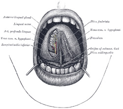 |
FIG. 1013– The mouth cavity. The apex of the tongue is turned upward, and on the right side a superficial dissection of its under surface has been made. (See enlarged image) |
| |
| The papillæ vallatæ (circumvallate papillæ) (Fig. 1015) are of large size, and vary from eight to twelve in number. They are situated on the dorsum of the tongue immediately in front of the foramen cecum and sulcus terminalis, forming a row on either side; the two rows run backward and medialward, and meet in the middle line, like the limbs of the letter V inverted. Each papilla consists of a projection of mucous membrane from 1 to 2 mm. wide, attached to the bottom of a circular depression of the mucous membrane; the margin of the depression is elevated to form a wall (vallum), and between this and the papilla is a circular sulcus termed the fossa. The papilla is shaped like a truncated cone, the smaller end being directed downward and attached to the tongue, the broader part or base projecting a little above the surface of the tongue and being studded with numerous small secondary papillæ and covered by stratified squamous epithelium. | 82 |
| The papillæ fungiformes (fungiform papillæ) (Fig. 1017), more numerous than the preceding, are found chiefly at the sides and apex, but are scattered irregularly and sparingly over the dorsum. They are easily recognized, among the other papillæ, by their large size, rounded eminences, and deep red color. They are narrow at their attachment to the tongue, but broad and rounded at their free extremities, and covered with secondary papillæ. | 83 |
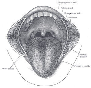 |
FIG. 1014– The mouth cavity. The cheeks have been slit transversely and the tongue pulled forward. (See enlarged image) |
| |
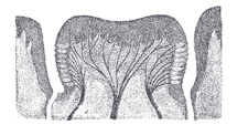 |
FIG. 1015– Circumvallate papilla in vertical section, showing arrangement of the taste-buds and nerves. (See enlarged image) |
| |
| The papillæ filiformes (filiform or conical papilæ) (Fig. 1016) cover the anterior two-thirds of the dorsum. They are very minute, filiform in shape, and arranged in lines parallel with the two rows of the papillæ vallatæ, excepting at the apex of the organ, where their direction is transverse. Projecting from their apices are numerous filamentous processes, or secondary papillæ these are of a whitish tint, owing to the thickness and density of the epithelium of which they are composed, which has here undergone a peculiar modification, the cells having become cornified and elongated into dense, imbricated, brush-like processes. They contain also a number of elastic fibers, which render them firmer and more elastic than the papillæ of mucous membrane generally. The larger and longer papillæ of this group are sometimes termed papillæ conicæ. | 84 |
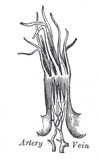 |
FIG. 1016– A filiform papilla. Magnified (See enlarged image) |
| |
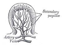 |
FIG. 1017– Section of a fungiform papilla. Magnified. (See enlarged image) |
| |
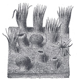 |
FIG. 1018– Semidiagrammatic view of a portion of the mucous membrane of the tongue. Two fungiform papillæ are shown. On some of the filiform papillæ the epithelial prolongations stand erect, in one they are spread out, and in three they are folded in. (See enlarged image) |
| |
| The papillæ simplices are similar to those of the skin, and cover the whole of the mucous membrane of the tongue, as well as the larger papillæ. They consist of closely set microscopic elevations of the corium, each containing a capillary loop, covered by a layer of epithelium. | 85 |
| |
| Muscles of the Tongue.—The tongue is divided into lateral halves by a median fibrous septum which extends throughout its entire length and is fixed below to the hyoid bone. In either half there are two sets of muscles, extrinsic and intrinsic; the former have their origins outside the tongue, the latter are contained entirely within it. | 86 |
| The extrinsic muscles (Fig. 1019) are: | 87 |
| Genioglossus. |
| Hyoglossus. |
| Chondroglossus. |
| Styloglossus.
| |
| | Glossopalatinus. 160 | 88 |
|
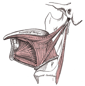 |
FIG. 1019– Extrinsic muscles of the tongue. Left side. (See enlarged image) |
| |
| The Genioglossus (Geniohyoglossus) is a flat triangular muscle close to and parallel with the median plane, its apex corresponding with its point of origin from the mandible, its base with its insertion into the tongue and hyoid bone. It arises by a short tendon from the superior mental spine on the inner surface of the symphysis menti, immediately above the Geniohyoideus, and from this point spreads out in a fan-like form. The inferior fibers extend downward, to be attached by a thin aponeurosis to the upper part of the body of the hyoid bone, a few passing between the Hyoglossus and Chondroglossus to blend with the Constrictores pharyngis; the middle fibers pass backward, and the superior ones upward and forward, to enter the whole length of the under surface of the tongue, from the root to the apex. The muscles of opposite sides are separated at their insertions by the median fibrous septum of the tongue; in front, they are more or less blended owing to the decussation of fasciculi in the median plane. | 89 |
| The Hyoglossus, thin and quadrilateral, arises from the side of the body and from the whole length of the greater cornu of the hyoid bone, and passes almost vertically upward to enter the side of the tongue, between the Styloglossus and Longitudinalis inferior. The fibers arising from the body of the hyoid bone overlap those from the greater cornu. | 90 |
| The Chondroglossus is sometimes described as a part of the Hyoglossus, but is separated from it by fibers of the Genioglossus, which pass to the side of the pharynx. It is about 2 cm. long, and arises from the medial side and base of the lesser cornu and contiguous portion of the body of the hyoid bone, and passes directly upward to blend with the intrinsic muscular fibers of the tongue, between the Hyoglossus and Genioglossus. | 91 |
| A small slip of muscular fibers is occasionally found, arising from the cartilago triticea in the lateral hyothyroid ligament and entering the tongue with the hindermost fibers of the Hyoglossus. | 92 |
| The Styloglossus, the shortest and smallest of the three styloid muscles, arises from the anterior and lateral surfaces of the styloid process, near its apex, and from the stylomandibular ligament. Passing downward and forward between the internal and external carotid arteries, it divides upon the side of the tongue near its dorsal surface, blending with the fibers of the Longitudinalis inferior in front of the Hyoglossus; the other, oblique, overlaps the Hyoglossus and decussates with its fibers. | 93 |
| The intrinsic muscles (Fig. 1020) are: | 94 |
| Longitudinalis superior. |
| Transversus. |
| Longitudinalis inferior. |
| Verticalis. |
|
| The Longitudinalis linguæ superior (Superior lingualis) is a thin stratum of oblique and longitudinal fibers immediately underlying the mucous membrane on the dorsum of the tongue. It arises from the submucous fibrous layer close to the epiglottis and from the median fibrous septum, and runs forward to the edges of the tongue. | 95 |
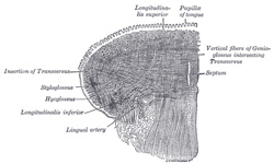 |
FIG. 1020– Coronal section of tongue, showing intrinsic muscles. (Altered from Krause.) (See enlarged image) |
| |
| The Longitudinalis linguæ inferior (Inferior lingualis) is a narrow band situated on the under surface of the tongue between the Genioglossus and Hyoglossus. It extends from the root to the apex of the tongue: behind, some of its fibers are connected with the body of the hyoid bone; in front it blends with the fibers of the Styloglossus. | 96 |
| The Transversus linguæ (Transverse lingualis) consists of fibers which arise from the median fibrous septum and pass lateralward to be inserted into the submucous fibrous tissue at the sides of the tongue. | 97 |
| The Verticalis linguæ (Vertical lingualis) is found only at the borders of the forepart of the tongue. Its fibers extend from the upper to the under surface of the organ. | 98 |
| The median fibrous septum of the tongue is very complete, so that the anastomosis between the two lingual arteries is not very free. | 99 |
| |
| Nerves.—The muscles of the tongue described above are supplied by the hypoglossal nerve. | 100 |
| |
| Actions.—The movements of the tongue, although numerous and complicated, may be understood by carefully considering the direction of the fibers of its muscles. The Genioglossi, by means of their posterior fibers, draw the root of the tongue forward, and protrude the apex from the mouth. The anterior fibers draw the tongue back into the mouth. The two muscles acting in their entirety draw the tongue downward, so as to make its superior surface concave from side to side, forming a channel along which fluids may pass toward the pharynx, as in sucking. The Hyoglossi depress the tongue, and draw down its sides. The Styloglossi draw the tongue upward and backward. The Glossopalatini draw the root of the tongue upward. The intrinsic muscles are mainly concerned in altering the shape of the tongue, whereby it becomes shortened, narrowed, or curved in different directions; thus, the Longitudinalis superior and inferior tend to shorten the tongue, but the former, in addition, turn the tip and sides upward so as to render the dorsum concave, while the latter pull the tip downward and render the dorsum convex. The Transversus narrows and elongates the tongue, and the Verticalis flattens and broadens it. The complex arrangement of the muscular fibers of the tongue, and the various directions in which they run, give to this organ the power of assuming the forms necessary for the enunciation of the different consonantal sounds; and Macalister states “there is reason to believe that the musculature of the tongue varies in different races owing to the hereditary practice and habitual use of certain motions required for enunciating the several vernacular languages.” | 101 |
| |
| Structure of the Tongue.—The tongue is partly invested by mucous membrane and a submucous fibrous layer. | 102 |
| The mucous membrane (tunica mucosa linguæ) differs in different parts. That covering the under surface of the organ is thin, smooth, and identical in structure with that lining the rest of the oral cavity. The mucous membrane of the dorsum of the tongue behind the foramen cecum and sulcus terminalis is thick and freely movable over the subjacent parts. It contains a large number of lymphoid follicles, which together constitute what is sometimes termed the lingual tonsil. Each follicle forms a rounded eminence, the center of which is perforated by a minute orifice leading into a funnel-shaped cavity or recess; around this recess are grouped numerous oval or rounded nodules of lymphoid tissue, each enveloped by a capsule derived from the submucosa, while opening into the bottom of the recesses are also seen the ducts of mucous glands. The mucous membrne on the anterior part of the dorsum of the tongue is thin, intimately adherent to the muscular tissue, and presents numerous minute surface eminences, the papillæ of the tongue. It consists of a layer of connective tissue, the corium or mucosa, covered with epithelium. | 103 |
| The epithelium is of the stratified squamous variety, similar to but much thinner than that of the skin: and each papilla has a separate investment from root to summit. The deepest cells may sometimes be detached as a separate layer, corresponding to the rete mucosum, but they never contain coloring matter. | 104 |
| The corium consists of a dense felt-work of fibrous connective tissue, with numerous elastic fibers, firmly connected with the fibrous tissue forming the septa between the muscular bundles of the tongue. It contains the ramifications of the numerous vessels and nerves from which the papillæ are supplied, large plexuses of lymphatic vessels, and the glands of the tongue. | 105 |
| Structure of the Papillæ.—The papillæ apparently resemble in structure those of the cutis, consisting of cone-shaped projections of connective tissue, covered with a thick layer of stratified squamous epithelium, and containing one or more capillary loops among which nerves are distributed in great abundance. If the epithelium be removed, it will be found that they are not simple elevations like the papillæ of the skin, for the surface of each is studded with minute conical processes which form secondary papillæ. In the papillæ vallatæ, the nerves are numerous and of large size; in the papillæ fungiformes they are also numerous, and end in a plexiform net-work, from which brush-like branches proceed; in the papillæ filiformes, their mode of termination is uncertain. | 106 |
| |
| Glands of the Tongue.—The tongue is provided with mucous and serous glands. | 107 |
| The mucous glands are similar in structure to the labial and buccal glands. They are found especially at the back part behind the vallate papillæ, but are also present at the apex and marginal parts. In this connection the anterior lingual glands (Blandin or Nuhn) require special notice. They are situated on the under surface of the apex of the tongue (Fig. 1013), one on either side of the frenulum, where they are covered by a fasciculus of muscular fibers derived from the Styloglossus and Longitudinalis inferior. They are from 12 to 25 mm. long, and about 8 mm. broad, and each opens by three or four ducts on the under surface of the apex. | 108 |
| The serous glands occur only at the back of the tongue in the neighborhood of the taste-buds, their ducts opening for the most part into the fossæ of the vallate papillæ. These glands are racemose, the duct of each branching into several minute ducts, which end in alveoli, lined by a single layer of more or less columnar epithelium. Their secretion is of a watery nature, and probably assists in the distribution of the substance to be tasted over the taste area. (Ebner.) | 109 |
| The septum consists of a vertical layer of fibrous tissue, extending throughout the entire length of the median plane of the tongue, though not quite reaching the dorsum. It is thicker behind than in front, and occasionally contains a small fibrocartilage, about 6 mm. in length. It is well displayed by making a vertical section across the organ. | 110 |
| The hyoglossal membrane is a strong fibrous lamina, which connects the under surface of the root of the tongue to the body of the hyoid bone. This membrane receives, in front, some of the fibers of the Genioglossi. | 111 |
| Taste-buds, the end-organs of the gustatory sense, are scattered over the mucous membrane of the mouth and tongue at irregular intervals. They occur especially in the sides of the vallate papillæ. In the rabbit there is a localized area at the side of the base of the tongue, the papilla foliata, in which they are especially abundant (Fig. 1021). They are described under the organs of the senses (page 991). | 112 |
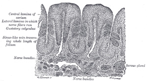 |
FIG. 1021– Vertical section of papilla foliata of the rabbit, passing across the folia. (Ranvier.) (See enlarged image) |
| |
| |
| Vessels and Nerves.—The main artery of the tongue is the lingual branch of the external carotid, but the external maxillary and ascending pharyngeal also give branches to it. The veins open into the internal jugular. | 113 |
| The lymphatics of the tongue have been described on page 696. | 114 |
| The sensory nerves of the tongue are: (1) the lingual branch of the mandibular, which is distributed to the papillæ at the forepart and sides of the tongue, and forms the nerve of ordinary sensibility for its anterior two-thirds; (2) the chorda tympani branch of the facial, which runs in the sheath of the lingual, and is generally regarded as the nerve of taste for the anterior two-thirds; this nerve is a continuation of the sensory root of the facial (nervus intermedius); (3) the lingual branch of the glossopharyngeal, which is distributed to the mucous membrane at the base and sides of the tongue, and to the papillæ vallatæ, and which supplies both gustatory filaments and fibers of general sensation to this region; (4) the superior laryngeal, which sends some fine branches to the root near the epiglottis. | 115 |
| |
| The Salivary Glands (Fig. 1024).—Three large pairs of salivary glands communicate with the mouth, and pour their secretion into its cavity; they are the parotid, submaxillary, and sublingual. | 116 |
| |
| Parotid Gland (glandula parotis).—The parotid gland (Figs. 1022, 1023), the largest of the three, varies in weight from 14 to 28 gm. It lies upon the side of the face, immediately below and in front of the external ear. The main portion of the gland is superficial, somewhat flattened and quadrilateral in form, and is placed between the ramus of the mandible in front and the mastoid process and Sternocleidomastoideus behind, overlapping, however, both boundaries. Above, it is broad and reaches nearly to the zygomatic arch; below, it tapers somewhat to about the level of a line joining the tip of the mastoid process to the angle of the mandible. The remainder of the gland is irregularly wedge-shaped, and extends deeply inward toward the pharyngeal wall. | 117 |
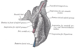 |
FIG. 1022– Right parotid gland. Posterior and deep aspects. (See enlarged image) |
| |
| The gland is enclosed within a capsule continuous with the deep cervical fascia; the layer covering the superficial surface is dense and closely adherent to the gland; a portion of the fascia, attached to the styloid process and the angle of the mandible, is thickened to form the stylomandibular ligament which intervenes between the parotid and submaxillary glands. | 118 |
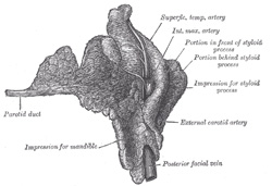 |
FIG. 1023– Right parotid gland. Deep and anterior aspects. (See enlarged image) |
| |
| The anterior surface of the gland is moulded on the posterior border of the ramus of the mandible, clothed by the Pterygoideus internus and Masseter. The inner lip of the groove dips, for a short distance, between the two Pterygoid muscles, while the outer lip extends for some distance over the superficial surface of the Masseter; a small portion of this lip immediately below the zygomatic arch is usually detached, and is named the accessory part (socia parotidis) of the gland. | 119 |
| The posterior surface is grooved longitudinally and abuts against the external acoustic meatus, the mastoid process, and the anterior border of the Sternocleidomastoideus. | 120 |
| The superficial surface, slightly lobulated, is covered by the integument, the superficial fascia containing the facial branches of the great auricular nerve and some small lymph glands, and the fascia which forms the capsule of the gland. | 121 |
| The deep surface extends inward by means of two processes, one of which lies on the Digastricus, styloid process, and the styloid group of muscles, and projects under the mastoid process and Sternocleidomastoideus; the other is situated in front of the styloid process, and sometimes passes into the posterior part of the mandibular fossa behind the temporomandibular joint. The deep surface is in contact with the internal and external carotid arteries, the internal jugular vein, and the vagus and glossopharyngeal nerves. | 122 |
| The gland is separated from the pharyngeal wall by some loose connective tissue. | 123 |
| |
| Structures within the Gland.—The external carotid artery lies at first on the deep surface, and then in the substance of the gland. The artery gives off its posterior auricular branch which emerges from the gland behind; it then divides into its terminal branches, the internal maxillary and superficial temporal; the former runs forward deep to the neck of the mandible; the latter runs upward across the zygomatic arch and gives off its transverse facial branch which emerges from the front of the gland. Superficial to the arteries are the superficial temporal and internal maxillary veins, uniting to form the posterior facial vein; in the lower part of the gland this vein splits into anterior and posterior divisions. The anterior division emerges from the gland and unites with the anterior facial to form the common facial vein; the posterior unites in the gland with the posterior auricular to form the external jugular vein. On a still more superficial plane is the facial nerve, the branches of which emerge from the borders of the gland. Branches of the great auricular nerve pierce the gland to join the facial, while the auriculotemporal nerve issues from the upper part of the gland. | 124 |
| The parotid duct (ductus parotideus; Stensen’s duct) is about 7 cm. long. It begins by numerous branches from the anterior part of the gland, crosses the Masseter, and at the anterior border of this muscle turns inward nearly at a right angle, passes through the corpus adiposum of the cheek and pierces the Buccinator; it then runs for a short distance obliquely forward between the Buccinator and mucous membrane of the mouth, and opens upon the oral surface of the cheek by a small orifice, opposite the second upper molar tooth. While crossing the Masseter, it receives the duct of the accessory portion; in this position it lies between the branches of the facial nerve; the accessory part of the gland and the transverse facial artery are above it. | 125 |
| |
| Structure.—The parotid duct is dense, its wall being of considerable thickness; its canal is about the size of a crow-quill, but at its orifice on the oral surface of the cheek its lumen is greatly reduced in size. It consists of a thick external fibrous coat which contains contractile fibers, and of an internal or mucous coat lined with short columnar epithelium. | 126 |
| |
| Vessels and Nerves.—The arteries supplying the parotid gland are derived from the external carotid, and from the branches given off by that vessel in or near its substance. The veins empty themselves into the external jugular, through some of its tributaries. The lymphatics end in the superficial and deep cervical lymph glands, passing in their course through two or three glands, placed on the surface and in the substance of the parotid. The nerves are derived from the plexus of the sympathetic on the external carotid artery, the facial, the auriculotemporal, and the great auricular nerves. It is probable that the branch from the auriculotemporal nerve is derived from the glossopharyngeal through the otic ganglion. At all events, in some of the lower animals this has been proved experimentally to be the case. | 127 |
| |
| Submaxillary Gland (glandula submaxillaris).—The submaxillary gland (Fig. 1024) is irregular in form and about the size of a walnut. A considerable part of it is situated in the submaxillary triangle, reaching forward to the anterior belly of the Digastricus and backward to the stylomandibular ligament, which intervenes between it and the parotid gland. Above, it extends under cover of the body of the mandible; below, it usually overlaps the intermediate tendon of the Digastricus and the insertion of the Stylohyoideus, while from its deep surface a tongue-like deep process extends forward above the Mylohyoideus muscle. | 128 |
| Its superficial surface consists of an upper and a lower part. The upper part is directed outward, and lies partly against the submaxillary depression on the inner surface of the body of the mandible, and partly on the Pterygoideus internus. The lower part is directed downward and outward, and is covered by the skin, superficial fascia, Platysma, and deep cervical fascia; it is crossed by the anterior facial vein and by filaments of the facial nerve; in contact with it, near the mandible, are the submaxillary lymph glands. | 129 |
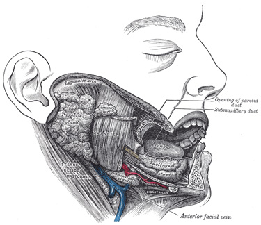 |
FIG. 1024– Dissection, showing salivary glands of right side. (See enlarged image) |
| |
| The deep surface is in relation with the Mylohyoideus, Hyoglossus, Styloglossus, Stylohyoideus, and posterior belly of the Digastricus; in contact with it are the mylohyoid nerve and the mylohyoid and submental vessels. | 130 |
| The external maxillary artery is imbedded in a groove in the posterior border of the gland. | 131 |
| The deep process of the gland extends forward between the Mylohyoideus below and externally, and the Hyoglossus and Styloglossus internally; above it is the lingual nerve and submaxillary ganglion; below it the hypoglossal nerve and its accompanying vein. | 132 |
| The submaxillary duct (ductus submaxillaris; Wharton’s duct) is about 5 cm. long, and its wall is much thinner than that of the parotid duct. It begins by numerous branches from the deep surface of the gland, and runs forward between the Mylohyoideus and the Hyoglossus and Genioglossus, then between the sublingual gland and the Genioglossus, and opens by a narrow orifice on the summit of a small papilla, at the side of the frenulum linguæ. On the Hyoglossus it lies between the lingual and hypoglossal nerves, but at the anterior border of the muscle it is crossed laterally by the lingual nerve; the terminal branches of the lingual nerve ascend on its medial side. | 133 |
| |
| Vessels and Nerves.—The arteries supplying the submaxillary gland are branches of the external maxillary and lingual. Its veins follow the course of the arteries. The nerves are derived from the submaxillary ganglion, through which it receives filaments from the chorda tympani of the facial nerve and the lingual branch of the mandibular, sometimes from the mylohyoid branch of the inferior alveolar, and from the sympathetic. | 134 |
| |
| Sublingual Gland (glandula sublingualis).—The sublingual gland (Fig. 1024) is the smallest of the three glands. It is situated beneath the mucous membrane of the floor of the mouth, at the side of the frenulum linguæ, in contact with the sublingual depression on the inner surface of the mandible, close to the symphysis. It is narrow, flattened, shaped somewhat like an almond, and weighs nearly 2 gm. It is in relation, above, with the mucous membrane; below, with the Mylohyoideus; behind, with the deep part of the submaxillary gland; laterally, with the mandible; and medially, with the Genioglossus, from which it is separated by the lingual nerve and the submaxillary duct. Its excretory ducts are from eight to twenty in number. Of the smaller sublingual ducts (ducts of Rivinus), some join the submaxillary duct; others open separately into the mouth, on the elevated crest of mucous membrane (plica sublingualis), caused by the projection of the gland, on either side of the frenulum linguæ. One or more join to form the larger sublingual duct (duct of Bartholin), which opens into the submaxillary duct. | 135 |
| |
| Vessels and Nerves.—The sublingual gland is supplied with blood from the sublingual and submental arteries. Its nerves are derived from the lingual, the chorda tympani, and the sympathetic. | 136 |
| |
| Structure of the Salivary Glands.—The salivary glands are compound racemose glands, consisting of numerous lobes, which are made up of smaller lobules, connected together by dense areolar tissue, vessels, and ducts. Each lobule consists of the ramifications of a single duct, the branches ending in dilated ends or alveoli on which the capillaries are distributed. The alveoli are enclosed by a basement membrane, which is continuous with the membrana propria of the duct and consists of a net-work of branched and flattened nucleated cells. | 137 |
| The alveoli of the salivary glands are of two kinds, which differ in the appearance of their secreting cells, in their size, and in the nature of their secretion. (1) The mucous variety secretes a viscid fluid, which contains mucin; (2) the serous variety secretes a thinner and more watery fluid. The sublingual gland consists of mucous, the parotid of serous alveoli. The submaxillary contains both mucous and serous alveoli, the latter, however, preponderating. | 138 |
| The cells in the mucous alveoli are columnar in shape. In the fresh condition they contain large granules of mucinogen. In hardened preparations a delicate protoplasmic net-work is seen, and the cells are clear and transparent. The nucleus is usually situated near the basement membrane, and is flattened. | 139 |
| In some alveoli are seen peculiar crescentic bodies, lying between the cells and the membrana propria. They are termed the crescents of Gianuzzi, or the demilunes of Heidenhain (Fig. 1025), and are composed of polyhedral granular cells, which Heidenhain regards as young epithelial cells destined to supply the place of those salivary cells which have undergone disintegration. This view, however, is not accepted by Klein. Fine canaliculi pass between the mucus-secreting cells to reach the demilunes and even penetrate the cells forming these structures. | 140 |
| In the serous alveoli the cells almost completely fill the cavity, so that there is hardly any lumen perceptible; they contain secretory granules imbedded in a closely reticulated protoplasm (Fig. 1026). The cells are more cubical than those of the mucous type; the nucleus of each is spherical and placed near the center of the cell, and the granules are smaller. | 141 |
| Both mucous and serous cells vary in appearance according to whether the gland is in a resting condition or has been recently active. In the former case the cells are large and contain many secretory granules; in the latter case they are shrunken and contain few granules, chiefly collected at the inner ends of the cells. The granules are best seen in fresh preparations. | 142 |
| The ducts are lined at their origins by epithelium which differs little from the pavement form. As the ducts enlarge, the epithelial cells change to the columnar type, and the part of the cell next the basement membrane is finely striated. | 143 |
| The lobules of the salivary glands are richly supplied with bloodvessels which form a dense net-work in the interalveolar spaces. Fine plexuses of nerves are also found in the interlobular tissue. The nerve fibrils pierce the basement membrane of the alveoli, and end in branched varicose filaments between the secreting cells. In the hilus of the submaxillary gland there is a collection of nerve cells termed Langley’s ganglion. | 144 |
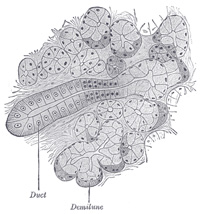 |
FIG. 1025– Section of submaxillary gland of kitten. Duct semidiagrammatic. X 200. (See enlarged image) |
| |
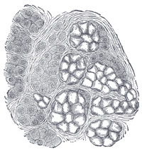 |
FIG. 1026– Human submaxillary gland. (R. Heidenhain.) At the right is a group of mucous alveoli, at the left a group of serous alveoli. (See enlarged image) |
| |
| |
| Accessory Glands.—Besides the salivary glands proper, numerous other glands are found in the mouth. Many of these glands are found at the posterior part of the dorsum of the tongue behind the vallate papillæ, and also along its margins as far forward as the apex. Others lie around and in the palatine tonsil between its crypts, and large numbers are present in the soft palate, the lips, and cheeks. These glands are of the same structure as the larger salivary glands, and are of the mucous or mixed type. | 145 |


