|
|
| Each lip of the valve is formed by a reduplication of the mucous membrane and of the circular muscular fibers of the intestine, the longitudinal fibers and peritoneum being continued uninterruptedly from the small to the large intestine. |
| The surfaces of the valve directed toward the ileum are covered with villi, and present the characteristic structure of the mucous membrane of the small intestine; while those turned toward the large intestine are destitute of villi, and marked with the orifices of the numerous tubular glands peculiar to the mucous membrane of the large intestine. These differences in structure continue as far as the free margins of the valve. It is generally maintained that this valve prevents reflux from the cecum into the ileum, but in all probability it acts as a sphincter around the end of the ileum and prevents the contents of the ileum from passing too quickly into the cecum. |
| The Colon is divided into four parts: the ascending, transverse, descending, and sigmoid. |
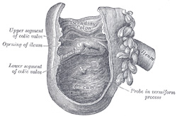 |
FIG. 1075– Interior of the cecum and lower end of ascending colon, showing colic valve. (See enlarged image)
|
| The Ascending Colon (colon ascendens) is smaller in caliber than the cecum, with which it is continuous. It passes upward, from its commencement at the cecum, opposite the colic valve, to the under surface of the right lobe of the liver, on the right of the gall-bladder, where it is lodged in a shallow depression, the colic impression; here it bends abruptly forward and to the left, forming the right colic (hepatic) flexure (Fig. 1056). It is retained in contact with the posterior wall of the abdomen by the peritoneum, which covers its anterior surface and sides, its posterior surface being connected by loose areolar tissue with the Iliacus, Quadratus lumborum, aponeurotic origin of Transversus abdominis, and with the front of the lower and lateral part of the right kidney. Sometimes the peritoneum completely invests it, and forms a distinct but narrow mesocolon. 1 It is in relation, in front, with the convolutions of the ileum and the abdominal parietes. |
| The Transverse Colon (colon transversum) the longest and most movable part of the colon, passes with a downward convexity from the right hypochondriac region across the abdomen, opposite the confines of the epigastric and umbilical zones, into the left hypochondriac region, where it curves sharply on itself beneath the lower end of the spleen, forming the left colic (splenic) flexure. In its course it describes an arch, the concavity of which is directed backward and a little upward; toward its splenic end there is often an abrupt U-shaped curve which may descend
|