| bone and undergo absorption. The inner alveolar border, usually described as arising from a separate ossific center (splenial center), is formed in the human mandible by an ingrowth from the main mass of the bone. At birth the bone consists of two parts, united by a fibrous symphysis, in which ossification takes place during the first year. |
| The foregoing description of the ossification of the mandible is based on the researches of Low 1 and Fawcett, 2 and differs somewhat from that usually given. |
Articulations.—The mandible articulates with the two temporal bones. |
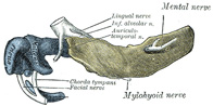 |
FIG. 178– Mandible of human embryo 24 mm. long. Outer aspect. (From model by Low.) (See enlarged image)
|
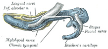 |
FIG. 179– Mandible of human embryo 24 mm. long. Inner aspect. (From model by Low.) (See enlarged image)
|
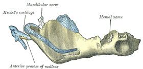 |
FIG. 180– Mandible of human embryo 95 mm. long. Outer aspect. Nuclei of cartilage stippled. (From model by Low.) (See enlarged image)
|
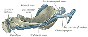 |
FIG. 181– Mandible of human embryo 95 mm. long. Inner aspect. Nuclei of cartilage stippled. (From model by Low.) (See enlarged image)
|
Changes Produced in the Mandible by Age—At birth (Fig. 182) the body of the bone is a mere shell, containing the sockets of the two incisor, the canine, and the two deciduous molar teeth, imperfectly partitioned off from one another. The mandibular canal is of large size, and runs near the lower border of the bone; the mental foramen opens beneath the socket of the first deciduous molar tooth. The angle is obtuse (175°), and the condyloid portion is nearly in line with the body. The coronoid process is of comparatively large size, and projects above the level of the condyle. |