| up in the fresh state by the interosseous talocalcaneal ligament. The posterior calcaneal articular surface is large and of an oval or oblong form. It articulates with the corresponding facet on the upper surface of the calcaneus, 1 and is deeply concave in the direction of its long axis which runs forward and lateralward at an angle of about 45° with the median plane of the body. The middle calcaneal articular surface is small, oval in form and slightly convex; it articulates with the upper surface of the sustentaculum tali of the calcaneus. |
| The medial surface presents at its upper part a pear-shaped articular facet for the medial malleolus, continuous above with the trochlea; below the articular surface is a rough depression for the attachment of the deep portion of the deltoid ligament of the ankle-joint. |
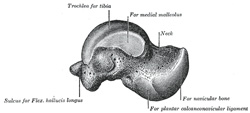 |
FIG. 272– Left talus, medial surface. (See enlarged image)
|
 |
FIG. 273– Left talus, lateral surface. (See enlarged image)
|
| The lateral surface carries a large triangular facet, concave from above downward, for articulation with the lateral malleolus; its anterior half is continuous above with the trochlea; and in front of it is a rough depression for the attachment of the anterior talofibular ligament. Between the posterior half of the lateral border of the trochlea and the posterior part of the base of the fibular articular surface is a triangular facet (Fawcett
2) which comes into contact with the transverse inferior tibiofibular ligament during flexion of the ankle-joint; below the base of this facet is a groove which affords attachment to the posterior talofibular ligament. |
| The posterior surface is narrow, and traversed by a groove running obliquely
|