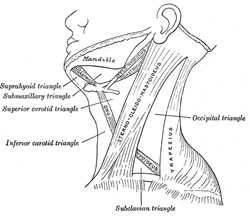
Anterior Triangle.—The anterior triangle is bounded, in front, by the middle line of the neck; behind, by the anterior margin of the Sternocleidomastoideus; its base, directed upward, is formed by the lower border of the body of the mandible, and a line extending from the angle of the mandible to the mastoid process; its apex is below, at the sternum. This space is subdivided into four smaller triangles by the Digastricus above, and the superior belly of the Omohyoideus below. These smaller triangles are named the inferior carotid, the superior carotid, the submaxillary, and the suprahyoid. |
| The Inferior Carotid, or Muscular Triangle, is bounded, in front, by the median line of the neck from the hyoid bone to the sternum; behind, by the anterior margin of the Sternocleidomastoideus; above, by the superior belly of the Omohyoideus. It is covered by the integument, superficial fascia, Platysma, and deep fascia, ramifying in which are some of the branches of the supraclavicular nerves. Beneath these superficial structures are the Sternohyoideus and Sternothyreoideus, which, together with the anterior margin of the Sternocleidomastoideus, conceal the lower part of the common carotid artery. 1 This vessel is enclosed within its sheath, together with the internal jugular vein and vagus nerve; the vein lies lateral to the artery on the right side of the neck, but overlaps it below on the left side; the nerve lies between the artery and vein, on a plane posterior to both.
|