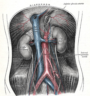
Anatomy of the Human Body > Page 602 CONTENTS · ILLUSTRATIONS · SUBJECT INDEX

  PREVIOUS NEXT  

behind the trachea and the esophagus, and may anastomose with the highest right intercostal artery. It represents the remains of the right dorsal aorta, and in a small proportion of cases is enlarged to form the first part of the right subclavian artery.
 
5a. 2. The Abdominal Aorta
 
  
(Aorta Abdominalis)


The abdominal aorta (Fig. 531) begins at the aortic hiatus of the diaphragm, in front of the lower border of the body of the last thoracic vertebra, and, descending in front of the vertebral column, ends on the body of the fourth lumbar vertebra, commonly a little to the left of the middle line, 1 by dividing into the two common iliac arteries. It diminishes rapidly in size, in consequence of the many large branches which it gives off. As it lies upon the bodies of the vertebræ, the curve which it describes is convex forward, the summit of the convexity corresponding to the third lumbar vertebra.


FIG. 531– The abdominal aorta and its branches. (See enlarged image)

Note 1.  Lord Lister, having accurately examined 30 bodies in order to ascertain the exact point of termination of this vessel, found it “either absolutely, or almost absolutely, mesial in 15, while in 13 it deviated more or less to the left, and in 2 was slightly to the right.” System of Surgery, edited by T. Holmes, 2d ed., v, 652. [back]

  PREVIOUS NEXT