Anatomy of the Human Body > Page 757 CONTENTS · ILLUSTRATIONS · SUBJECT INDEX

  PREVIOUS NEXT  

S2, S3 and S4 (Bruce). 1 Behind it is a dorso-medial column of small cells, which is not represented in L5, S1, S2 nor below S4. Its axons probably pass into the dorsal rami of the spinal nerves to supply the dorsal musculature of the spinal column. In the cervical and lumbar enlargements, where the anterior column is expanded in a lateral direction, the following additional columns are present, viz.: (a) antero-lateral, which consists of two groups, one in C4, C5, C6 the other in C6, C7, C8 in the cervical enlargement and of a group from L2 to S2 in the lumbo-sacral enlargement; (b) postero-lateral, in the lower five cervical, lower four lumbar, and upper three sacral segments; (c) post-postero-lateral, in the last cervical, first thoracic, and upper three sacral segments; and (d) a central, in the lower four lumbar and upper two sacral segments. These cell groups are evidently related to the nerve roots of the brachial and sacral plexuses and supply fibers to the muscles of the arm and leg. Throughout the base of the anterior column are scattered solitary cells, the axons of some of which form crossed commissural fibers, while others constitute the motor fibers of the posterior nerve roots. (See footnote, page 755.)


FIG. 671– Transverse sections of the medulla spinalis at different levels to show the arrangement of the principal cell columns. (See enlarged image)


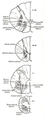
Nerve Cells in the Lateral Column.—These form a column which is best marked where the lateral gray column is differentiated, viz., in the thoracic region; 2 but it can be traced throughout the entire length of the medulla spinalis in the form of groups of small cells which are situated in the anterior part of the formatio reticularis. In the upper part of the cervical region and lower part of the medulla oblongata as well as in the third and fourth sacral segments this column is again differentiated. In the medulla it is known as the lateral nucleus. The cells of this column are fusiform or star-shaped, and of a medium size: the axons of some of them pass into
Note 1.  Topographical Atlas of the Spinal Cord, 1901. [back]
Note 2.  According to Bruce and Pirie (B. M> J., November 17, 1906) this column extends from the middle of the eighth cervical segment to the lower part of the second lumbar or the upper part of the third lumbar segment. [back]

  PREVIOUS NEXT